Salter-Harris Typ 2 Fraktur


Salter-Harris
type II fracture • Salter-Harris type II fracture - Ganzer Fall bei Radiopaedia

Salter-Harris
classification • Salter-Harris fracture of apophysis - Ganzer Fall bei Radiopaedia

Salter-Harris
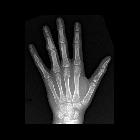
classification • Salter-Harris type II fracture - fifth finger - Ganzer Fall bei Radiopaedia

Salter-Harris
type II fracture • Salter-Harris type II fracture - proximal phalanx - Ganzer Fall bei Radiopaedia

Salter-Harris
type II fracture • Salter-Harris type II radial head fracture - Ganzer Fall bei Radiopaedia

Salter-Harris
type II fracture • Salter-Harris type II fracture - Ganzer Fall bei Radiopaedia

Salter-Harris
type II fracture • Salter-Harris type II fracture: proximal phalanx - Ganzer Fall bei Radiopaedia

Salter-Harris
type II fracture • Salter-Harris type II fracture: finger - Ganzer Fall bei Radiopaedia

Salter-Harris
type II fracture • Salter-Harris type II fracture: proximal phalanx - Ganzer Fall bei Radiopaedia

Salter-Harris
classification • Ankle fracture - Salter-Harris type II - Ganzer Fall bei Radiopaedia

Salter-Harris
type II fracture • Salter-Harris type II fracture - wrist - Ganzer Fall bei Radiopaedia

Salter-Harris
type II fracture • Salter-Harris type II fracture - Ganzer Fall bei Radiopaedia

Salter-Harris
type II fracture • Salter-Harris type II fracture - Ganzer Fall bei Radiopaedia

Salter-Harris
type II fracture • 5th toe fracture - Salter-Harris type II - Ganzer Fall bei Radiopaedia

Salter-Harris
type II fracture • Salter-Harris type II fracture - Ganzer Fall bei Radiopaedia

Salter-Harris
classification • Salter-Harris illustrations - Ganzer Fall bei Radiopaedia

School ager
who stubbed their first toe and now has pain there. AP (left) and lateral (right) radiographs of the first toe show soft tissue swelling around the toe and a small bone fragment in the center of the distal physis anteriorly.The diagnosis was a non-displaced Salter-Harris Type II fracture of the distal phalanx of the first toe.
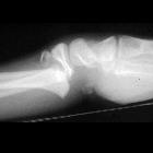
Teenager with
wrist pain after a fall. Lateral radiograph of the wrist shows a fracture through the radial physis and extending up into the radial metaphysis.The diagnosis was a Salter-Harris Type II fracture of the radius.

Salter-Harris
II (Aitken-I-Fraktur) der distalen Tibia mit Hervorhebung des metaphysären Fragments, welches mit der Epiphyse verbunden bleibt, so genanntes Thurstan Holland-Fragment.
Salter-Harris type II fractures are the most common type of physeal fractures that occur in children. There is a fracture that extends through the physis and into a portion of the metaphysis. A triangular metaphyseal fragment, otherwise known as the Thurston Holland fragment, will be left intact.
Epidemiology
Approximately 75% of physeal fractures will be a Salter-Harris type II with 33-50% occurring at the distal radius. Other common fracture sites are the distal tibia, distal fibula and, phalanges .
Radiographic features
In reality, the majority of fractures that involve the physis have at least a small fragment of metaphysis associated with them and are therefore type II injuries.
Plain radiograph
- fracture through the physis
- Thurston Holland fragment
- angulation, displacement and rotation may occur
Siehe auch:


 Assoziationen und Differentialdiagnosen zu Salter-Harris Typ 2 Fraktur: