Schwannom des Nervus suprascapularis

Peripheral
schwannoma mimicking a bone tumour. Well-defined lytic lesion affecting part of the corpus, the neck and the glenoid of the scapula. Narrow transition zone with thin sclerotic rim is shown.
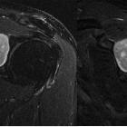
Peripheral
schwannoma mimicking a bone tumour. Hyperintense, well-defined lesion on T2-WI
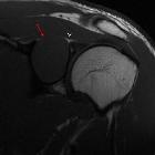
Peripheral
schwannoma mimicking a bone tumour. Isointense well-defined mass with epicentre in the spinoglenoid notch (red arrow). Note the close relationship with the suprascapular nerve (white arrow).
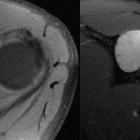
Peripheral
schwannoma mimicking a bone tumour. The lesion shows intense and homogenous enhancement after the administration of Gadolinium. Note the preserved rim of very thin cortical rest (arrow).
Schwannom des Nervus suprascapularis
Siehe auch:


 Assoziationen und Differentialdiagnosen zu Schwannom des Nervus suprascapularis: