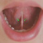
Speichelstein Ductus submandibularis

Imaging of
the sublingual and submandibular spaces. Sialolithiasis with resultant sialadenitis. Axial contrast-enhanced CT images at the level of the submandibular gland demonstrate two well-circumscribed calcifications in the distal right submandibular duct (a, arrow). The right submandibular duct is dilated proximal to the stones (b, arrowhead). There is enlargement and enhancement of the right submandibular gland (*) compared to the normal left submandibular gland

Imaging of
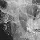
the sublingual and submandibular spaces. Sialolithiasis. a Conventional sialogram demonstrates multiple filling defects within Wharton’s (submandibular) duct, consistent with sialoliths. b, c In another patient, pre- and post-contrast injection sialogram images demonstrate duct obstruction at the site of a large stone (no contrast is present proximal to the stone, see arrow)

Sialolithiasis
vor allem linke Glandula submandibularis; im Volumen Rendering zeigen sich auch in der Glandula submandibularis rechts und kleine in der Glandula parotis Speichelsteine.
Speichelstein Ductus submandibularis
Siehe auch:
und weiter:

 Assoziationen und Differentialdiagnosen zu Speichelstein Ductus submandibularis:


