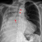
spindle cell carcinoma of lung

Surgical
resection of a rapidly growing pulmonary spindle cell carcinoma by robot-assisted thoracoscopic surgery: a case report. A Chest computed tomography (CT) scan shows a lobular mass of 33 mm located in the right upper lobe and B lymph node enlargement in station #4R. C Chest CT scan, taken 1 month after consultation, shows that the tumor diameter increased to 52 mm and D lymph node enlargement in station #4R

Surgical
resection of a rapidly growing pulmonary spindle cell carcinoma by robot-assisted thoracoscopic surgery: a case report. A 18F-fluorodeoxyglucose (FDG) positron emission tomography scan shows FDG accumulation with a maximum standardized uptake value (SUVmax) of 11.78 in the mass and B FDG accumulation with a SUVmax of 3.48 in the right lower paratracheal lymph node (station #4R)
Spindle cell carcinomas of the lung correspond to a very rare type of non-small cell lung carcinomas (NSCLC) classified under sarcomatoid carcinomas of the lungs.
Terminology
Although, in the past, some authors also used the term synonymously with pleomorphic carcinoma of the lung,since the 1999 version of the WHO classification of tumors, spindle cell carcinomas of the lung are considered as a separate entity.
Pathology
Tumors usually show a concurrent presence of malignant epithelial and homologous sarcomatoid spindle cell components by co-expressing cytokeratin and vimentin in various degrees.
Radiographic features
We do not know of any specific imaging features specifically for this tumor. The lesion may either present as endobronchial lesions or peripheral parenchymal lesions.
Siehe auch:
und weiter:

 Assoziationen und Differentialdiagnosen zu spindle cell carcinoma of lung:

