Stein der Nasennebenhöhlen

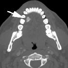
A rare case
of sinolith in the ethmoid sinus. Axial and coronal CT images reveal a calcified, hyperdense irregularly-shaped mass in the left ethmoid region bulging into the sphenoid sinus. Inflammatory tissue in bilateral ethmoidal air cells was also noted.

A rare case
of sinolith in the ethmoid sinus. Axial and coronal CT images reveal a calcified, hyperdense irregularly-shaped mass in the left ethmoid region bulging into the sphenoid sinus. Inflammatory tissue in ethmoidal air cells was also noted.

A rare case
of sinolith in the ethmoid sinus. MRI shows a signal void in the left ethmoid region on both T1 and T2 weighted axial images. Also inflammatory tissue in ethmoid air calls is demonstrated.

A rare case
of sinolith in the ethmoid sinus. MRI shows a signal void in the left ethmoid sinus on both T1 and T2 weighted axial images. Also inflammatory tissue in ethmoidal air cells is demonstrated.
Stein der Nasennebenhöhlen
Siehe auch:


 Assoziationen und Differentialdiagnosen zu Stein der Nasennebenhöhlen: