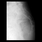
Steißbeinfraktur

Coccygeal fractures are generally low-severity injuries, which nonetheless can be diagnostically challenging. Diagnosis may be delayed or missed due to coccygeal anatomy and patient/technical factors (e.g. obesity, overlying bowel gas/feces).
Given that management of coccygeal fractures is nearly always non-operative, some radiology literature suggests that x-ray evaluation for coccygodynia is a waste of resources and exposes patients to unnecessary ionizing radiation, without having measurable impact on clinical outcome .
Pathology
Coccygeal fractures in younger adults tend to be after high-energy trauma. In elderly patients, these can represent insufficiency-type fractures .
Classification
Within the AO classification system, coccygeal fractures are classified as a subset of the sacrococcygeal fractures (classification A1).
Radiographic features
Most coccygeal fractures have a transverse orientation . Displacement of the fracture fragment is variable.
Plain radiograph
- best demonstrated on the lateral projection
Treatment and prognosis
As a rule, coccygeal fracture/dislocations are treated with non-operative management (e.g. cushioning and analgesia). Significant angulation or displacement may require closed reduction, often intra-anal manipulation.
Surgery is generally reserved for open injuries requiring soft tissue debridement, or chronic symptomatic injuries. In these cases, the fractured component is generally resected (coccygectomy).
Siehe auch:

