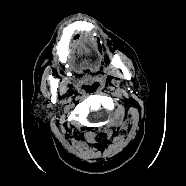
stensen's duct

The parotid duct, also known as Stensen duct, drains saliva from the parotid gland into the oral cavity. It primarily secretes serous saliva.
Gross anatomy
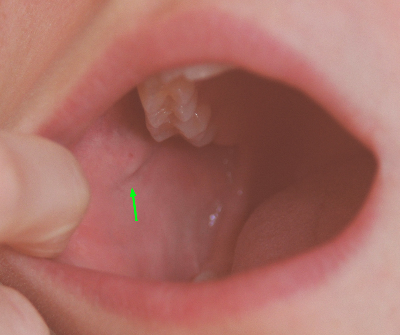
The parotid duct passes anteriorly through the buccal fat superficial to the masseter muscle and over its anterior border, then through the buccopharyngeal fascia and the buccinator muscle. It then continues between the buccal mucosa and the buccinator to its opening in the vestibule of the mouth, located next to the upper second molar tooth at the parotid ampulla. The buccinator muscle ensures that ballooning of the duct does not occur during blowing.
It is approximately 5 cm long, At four different points along its length, it has mean diameters ranging between 1.4 mm and 0.5 mm, with a maximum of 2.3 mm and a minimum of 0.1 mm, depending on the site .
Relations
Several other structures run alongside the parotid duct:
- superiorly: transverse facial artery
- inferiorly: buccal nerve
History and etymology
It is named after the Danish anatomist Niels Stensen (1638-1686) (also known as Nicolaus Steno) who was the first to describe it, initially in a sheep, in 1660. His colleague Sylvius (1614-1672) confirmed its presence in the human body and van Horne in Leyden named it after Stensen .
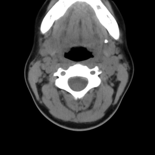
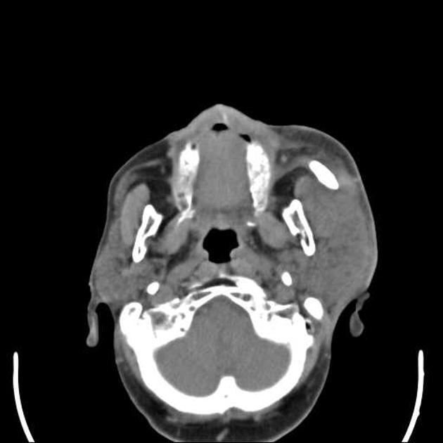
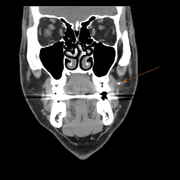
Related pathology
Blockage of the parotid duct can occur secondary to salivary duct stones or external compression. Either cause of obstruction can cause pain and parotitis. Stones are more common in the submandibular gland and duct.
Siehe auch:
- Glandula parotidea
- Sialolithiasis
- duct of Rivinus
- ducts of the salivary glands
- Wharton-Kanal
- parotid duct strictures
und weiter:


 Assoziationen und Differentialdiagnosen zu Ductus parotideus: