Stressfraktur des Talus

Bone stress
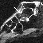
injury of the ankle in professional ballet dancers seen on MRI. Typical appearance of talar bone marrow edema observed on MR images of nine out of twelve ankles of symptomatic professional ballet dancers: Sagittal STIR (TR/TE = 1800/24) image of a 24-year-old male ballet dancer shows associated edema-type high signal within the talus (arrow).

Bone stress
injury of the ankle in professional ballet dancers seen on MRI. Sagittal STIR MR image of a 32-year-old female shows patchy edema signal within the talar neck (arrow).

Bone stress
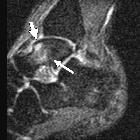
injury of the ankle in professional ballet dancers seen on MRI. Sagittal STIR MR image of a 25-year-old male shows patchy edema signal within the body of the talus (long arrow), which extends to the subchondral region of the talar dome (short arrow). This same edema pattern was observed in nine of the twelve ankles and may be related to chronic repetitive stress.

Bone stress
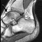
injury of the ankle in professional ballet dancers seen on MRI. Talar bone marrow edema in a symptomatic 24-year-old male (same subject as in figure 1). Sagittal T1-weighted (TR/TE = 380/18) MR image shows patchy low signal within the talus (arrow).

Stress
fracture of the posterior talar process in a female long-distance runner treated by osteosynthesis with screw fixation via two-portal hindfoot endoscopy: a case report. Preoperative non-contrast CT scan. a Sagittal and (b) axial views showing the fracture line located just lateral to the groove for flexor hallucis longus (FHL) tendon at a level just proximal to the subtalar joint. Posterior process of the talus comprising the medial (dotted arrow) and lateral (arrow) tubercles, separated by the groove for the FHL tendon. CT, computed tomography
Stressfraktur des Talus
Siehe auch:

 Assoziationen und Differentialdiagnosen zu Stressfraktur des Talus:
