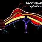
subperiostaler Abszess

School ager
with left eye swelling Axial CT with contrast of the orbits shows opacification of the left ethmoid sinus and bilateral sphenoid sinuses along with pre and post-septal inflammation of the left orbit. A lenticular fluid collection with rim enhancement is present along the medial wall of the left orbit.The diagnosis was orbital cellulitis with a subperiosteal abscess.

Lesions
involving the outer surface of the bone in children: a pictorial review. Septic cortical osteitis with subperiosteal abscess formation in a 7-year-old girl. Antero-posterior radiograph of the left tibia a shows an ill-defined lucency at the lateral aspect of the proximal tibial metaphysis (arrow). Axial STIR image b reveals a crescentic, hyperintense fluid collection adjacent to the proximal tibia at this level. This collection is hypointense with peripheral enhancement as seen on the post-contrast axial c and sagittal d images. Enhancement can also be seen through surrounding soft tissues
subperiostaler Abszess
Siehe auch:
- subperiostaler Abszess der Orbita
- subperiostales Hämatom
- subperiosteal abscess (mastoid)
- Pott's puffy tumour
- subperiostaler temporaler Abszess vom Ohr ausgehend
und weiter:

 Assoziationen und Differentialdiagnosen zu subperiostaler Abszess:

