superficial cerebral vein thrombosis


Cortical vein thrombosis, also known as superficial cerebral vein thrombosis, is a subset of cerebral venous thrombosis involving the superficial cerebral veins besides the dural sinus, often coexisting with deep cerebral vein thrombosis or dural venous sinus thrombosis. It has different clinical presentations relying on which segment is involved. Isolated cortical vein thrombosis without sinus involvement is reported to be extremely rare.
As such please refer to the cerebral venous thrombosis article for a general discussion.
Clinical presentation
It has a nonspecific pattern, ranging from essentially asymptomatic to coma and death, depending on the extension, collateral flow and association with deep cerebral vein thrombosis and dural venous sinus thrombosis. Please refer to the generic article for a broad discussion on clinical presentation: cerebral venous thrombosis.
Pathology
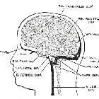
It is important to emphasize that cortical veins are extremely variable in number, size, and location . The cortical cerebral veins considered for this pathology are:
- superficial middle cerebral vein
- inferior anastomotic vein (vein of Labbe)
- superior anastomotic vein (vein of Trolard)
- small (unnamed) cortical veins
Venous infractions will be directly related to the presence or not of a collateral outflow.
Please refer to the generic article for a broad discussion on pathology and risk factors: cerebral venous thrombosis.
Radiographic features
Unenhanced CT is usually the first imaging investigation performed given the nonspecific clinical presentation in these cases. Unfortunately however CT is usually normal . Potential findings include:
- cord sign (MPR views)
- dense vein sign
- cortical edema
- cortical or peripheral venous hemorrhage (linear cortical density or gyriform heterogeneous hemorrhage)
Contrast-enhanced CT, especially with MPR CT venogram, may demonstrate a filling defect in a superficial vein or in a contiguous venous sinus. Focal gyral enhancement may occur.
MRI is more sensitive, but only when there is occlusion of the largest cortical veins. Occlusion involving small veins at the cortical level is difficult to identify .
Treatment and prognosis
For general discussion on treatment please refer to the parent article: cerebral venous thrombosis.
Siehe auch:

 Assoziationen und Differentialdiagnosen zu superficial cerebral vein thrombosis:


