Surgical hemostatic material


 nicht verwechseln mit: Gossypibom
Surgical hemostatic material is used to control bleeding intraoperatively and is hence frequently intentionally left in the operative bed, not to be confused with a gossypiboma which is caused by foreign material left behind in error. Its use has increased with the advent of minimally invasive surgery and compliments other hemostatic techniques such as cautery. It can mimic an abscess, tumor, lymph node or retained foreign body on imaging studies. Various types are available, the most common type being composed of oxidised regenerated cellulose. It may be deployed in a number of different forms, such as sheets, sponges, or powder.
Surgicel
Oxidized regenerated cellulose or Surgicel (Ethicon, part of Johnson & Johnson) is a bioabsorbable sterile knitted fabric prepared by the controlled oxidation of regenerated cellulose.
The mechanism of action whereby Surgicel® accelerates clotting is not completely understood, the effect appears to be physical rather than an alteration to the physiological clotting mechanism. The material can act as a scaffold to facilitate clot formation.
Surgicel® combines with blood into a brownish gelatinous mass which aids in the formation of a clot, as it is bioabsorbable it can be left in the surgical bed with almost no local tissue reaction. Retained Surgicel® is normally reabsorbed within 7-14 days.
Surgicel can absorb a large amount of blood and fluid, expanding in volume as it does so, with potential deleterious effects on surrounding structures . However, this effect is less pronounced within the abdominal cavity.
Differentiating hemostatic material from abscess
The most important factor is accurate clinical information on the use of surgical hemostatic material. If in doubt, discuss with the operating surgeon who should be able to advise whether surgical hemostatic material was used.
There are a number of appearances that can help to differentiate between surgical hemostatic material and an abscess :
- gas pockets are packed tightly, not discrete, and arranged in a linear fashion
- gas-fluid levels, commonly seen in an abscess, are usually not seen with surgical hemostatic material
- rim enhancement is generally not seen around surgical hemostatic material (although may occur after a few days due to a granulomatous reaction at the edges)
- surgical hemostatic material ‘collections’ tend to be organized in geometric shapes
Radiographic features
Ultrasound
Oxidised cellulose is seen as an echogenic mass with posterior reverberation artifact from the gas, mimicking a gas-containing abscess . There may be surrounding free fluid.
CT
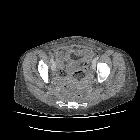
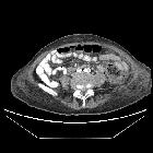
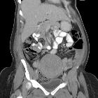
A well-defined "collection" comprised of mixed gas and soft tissue is seen, often surrounded by free fluid and adjacent to other collections. The gas bubbles are arranged in linear patterns, and the collection has a geometric shape. An enhancing margin typical of abscesses is usually not present.
Practical points
Be cautious about labeling well-defined clusters of admixed gas and soft tissue without definable marginal enhancement as abscesses. Check with the operating surgeon whether surgical hemostatic material was used.
See also

