thoracic actinomycosis infection

Thoracic actinomycosis refers to an uncommon indolent infection caused principally by the genus Actinomyces (higher prokaryotic bacteria belonging to the family Actinomyceataceae).
Epidemiology
While it is rare in general, the thoracic form actinomycosis constitutes ∼15% of the total burden of actinomycosis.
Clinical presentation
Most patients may initially experience a non productive cough and low-grade fever, but pulmonary complaints can be minimal. Spread to the pleura and chest wall may result in chest wall pain. The disease may present as a chronic debilitating illness. Some patients may have hemoptysis . Definitive diagnosis on clinical grounds can be difficult.
Pathology
Thoracic involvement usually results from aspiration of oropharyngeal or gastrointestinal secretions into the respiratory tract. Actinomyces israelii can be commonly found in the oral cavity (especially in those with poor oral hygiene or from extension of cervicofacial infections), and is thought to be responsible for the majority of pulmonary actinomyces infections. Actinomyces naeslundii is also present in normal oral flora, although it much less commonly associated with lung disease.
Since the condition can mimic a range of other pathologies on clinical and radiological grounds, tissue diagnosis is considered essential.
Distribution
There may be a peripheral and/or lower lobe predominance which probably reflects the role of aspiration in its pathogenesis . Most lesions may be unilateral .
Radiographic features
General
Thoracic actinomycosis can have variety of radiographic presentations which can also depend on time since infection.
According to Kim et al. at least four different forms have been described :
- lung parenchymal actinomycosis / pulmonary actinomycosis - is probably the commonest form
- bronchiectatic form of actinomycosis -
- secondary actinomycotic infection can involve a devitalized lobe or segment that already has been damaged by previous tuberculosis or by other bacterial infections which has lead to pre-existing bronchiectasis
- endobronchial actinomycosis associated with broncholithiasis - rare
- endobronchial actinomycosis associated with a foreign body - rare
Many of the features listed below pertain to pulmonary actinomycosis +/- its complications
Chest radiograph
While being non specific, The most common chest radiographic finding tends to be consolidation (usually non-segmental pneumonia in the lower zones and peripherally crossing fissures), or mass like lesions .
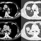
CT - HRCT chest
CT manifestations are also varied with each feature being non specific on its own. These include
- patchy air-space consolidation - relatively common feature
- may involve pleura and chest wall leading to empyema necessitans.
- dense consolidation may give air-bronchograms
- may have associated central areas of low attenuation .
- multifocal nodular appearances
- cavitation
- associated pleural thickening - relatively common feature
- pleural effusions - relatively common feature ~ 50%
- hilar, and/or mediastinal lymphadenopathy
Treatment and prognosis
A commonly accepted principal treatment of actinomycosis comprises of long-term administration of a high-dose intravenous antibiotic such as penicillin. Several recent studies have reported that the short-term treatment has also be successful .
Differential diagnosis
The imaging differential diagnosis can be wide ranging ranging on the type of manifestations. Therefore it may be more meaningful so consider a differential for each radiographic feature.
Siehe auch:
und weiter:


 Assoziationen und Differentialdiagnosen zu thorakale Aktinomykose: