tibial osteochondroma


Kartilaginäre
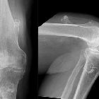
Exostosen an Femur, Tibia und Fibula. Synostose zwischen Tibia und Fibula. Röntgenbild ap.

Kartilaginäre
Exostosen an Femur, Tibia und Fibula. Synostose zwischen Tibia und Fibula. Röntgenbild seitlich


Lesions
involving the outer surface of the bone in children: a pictorial review. Benign solitary osteochondroma of the tibia in a 15-year-old boy. Lateral radiograph of the knee a shows a bony protrusion arising off the posterior aspect of the proximal tibial metaphysis; note the continuity with the medullary cavity of the underlying tibia (arrow). Sagittal b and axial c T2 MR images show a markedly hyperintense, thin cartilage cap surrounding this bony exostosis; an axial T1 post contrast MR image d reveals a thin, peripherally enhancing cartilage cap (arrow)

Typische
kartilaginäre Exostose mediale proximale Tibia mit breiter Basis und kontinuierlicher Fortsetzung der Spongiosa in die Exostose.


 Assoziationen und Differentialdiagnosen zu kartilaginäre Exostosen der Tibia:

