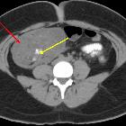
torquierte Dermoidzyste des Ovars

Mature cystic
teratoma with torsion. A Lobulated lesion (red arrow) is seen in the right lower abdomen with evidence of fat and calcification (tooth-like)(yellow arrow) within.

Mature cystic
teratoma with torsion. A Lobulated lesion (red arrow) is seen in the right lower abdomen with evidence of fat and calcification (tooth like)(yellow arrow) within.

Mature cystic
teratoma with torsion. A lobulated lesion (red arrow) is seen in the right lower abdomen and pelvis with evidence of fat and calcification (tooth-like)(yellow arrow) within the lesion.

Mature cystic
teratoma with torsion. A lobulated lesion (red arrow) is seen in the right lower abdomen and pelvis with evidence of fat and calcification (tooth-like)(yellow arrow) within the lesion.

Mature cystic
teratoma with torsion. A Lobulated lesion (red arrow) is seen on the right side in the pelvis. Enlarged left adnexa (blue arrow) is seen with twisted pedicle and midline displacement. Uterus is deviated to the right (green arrow).

Mature cystic
teratoma with torsion. Lesion (red arrow) in the right lower abdomen and pelvis with fat and calcification (yellow arrow). Enlarged left adnexa (blue arrow) with twisted pedicle and midline displacement. Uterus deviated to the right(green arrow).

Mature cystic
teratoma with torsion. Lesion (red arrow) with fat and calcification (yellow arrow) within. Minimal free fluid is seen inferior to the lesion (orange arrow). Note right side displacement of uterus(green arrow) and enlarged left adnexa (blue arrow).
torquierte Dermoidzyste des Ovars
Siehe auch:

 Assoziationen und Differentialdiagnosen zu torquierte Dermoidzyste des Ovars:
