Tubenruptur

Ruptured
ectopic pregnancy. Axial post-contrast MDCT revealed a pelvic rounded cyst-like structure with peripheral enhancement due to the extrauterine gestational sac (white arrow).

Imaging for
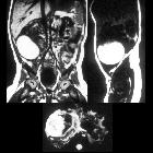
acute pelvic pain in pregnancy. A 27-year-old woman presenting at the emergency department with acute pelvic pain. Abdominal US (a) shows a rounded lesion adjacent to the uterus, clearly separate from the left ovary. Axial CT scan shows a voluminous mass in the pelvic cavity (b); for the characterisation an MRI was required. Axial (c) and coronal (d) T2-weighted and T1-weighted images (e) of the pelvis show a right heterogeneous adnexal mass (arrow) with fallopian tube haematoma. Note the normal ovary (short arrow) in (c). Pre-contrast T1-weighted fat-saturated image (f) shows bloody ascites (short arrows). These findings are due to ectopic pregnancy with tubal rupture and haemoperitoneum
Fallopian tube rupture is most often a complication of a tubal ectopic pregnancy where the pregnancy breaks open due to progressive growth. It can potentially lead to shock.
Pathology
Risk factors
Factors that raise the risk for a tubal rupture in a given tubal ectopic pregnancy include :
- history of pelvic surgery: tubal injury
- ovulation induction
- bilateral tubal ligation
- history of pelvic inflammatory disease
- previous ectopic pregnancy
- intrauterine contraceptive device use
- high beta-HCG levels: >5,000 mIU/ml
Radiographic features
Ultrasound
Can show a large amount of free peritoneal fluid and/or hemoperitoneum.
Siehe auch:
- tubal ectopic pregnancy
- Ovarialtorsion
- Intrauterinpessar
- Hydrosalpinx
- Morgagni-Hydatide
- Unterleibsentzündung
- Tubentorsion
- haematosalpinx
- tubal spasm
und weiter:

 Assoziationen und Differentialdiagnosen zu Tubenruptur:


