ureteral diverticula

Ureteral
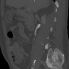
diverticula in a patient with recurrent urinary infections. Oblique MIP image shows two diverticula of the upper tract of the right ureter and of the middle tract of the left ureter.

Ureteral
diverticula in a patient with recurrent urinary infections. Axial MIP image shows the right ureteral diverticulum, with its little neck of connection to the ureteral lumen (arrow).

Ureteral
diverticula in a patient with recurrent urinary infections. Sagittal-oblique MIP image shows the right ureteral diverticulum, with its little neck of connection to the ureteral lumen.

Ureteral
diverticula in a patient with recurrent urinary infections. Coronal-oblique MIP image shows the left ureteral diverticulum, with its little neck of connection to the ureteral lumen.

Ureteral
diverticula in a patient with recurrent urinary infections. Volume rendering reconstruction accurately shows the two ureteral diverticula.
echte Ureterdivertikel
ureteral diverticula
Siehe auch:

 Assoziationen und Differentialdiagnosen zu echte Ureterdivertikel:

