verkalkte tracheale Noduli

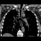
Tracheobronchopathia
osteochondroplastica. Cross sectional CT image showing diffuse submucosal calcified nodules of the anterolateral walls of the trachea, excluding the posterior membrane

Tracheobronchopathia
osteochondroplastica. Cross sectional CT image showing diffuse submucosal calcified nodules of the anterolateral walls of the trachea, excluding the posterior membrane

Tracheobronchopathia
osteochondroplastica. Cross sectional CT image showing diffuse submucosal calcified nodules of the anterolateral walls of the trachea, excluding the posterior membrane

Tracheobronchopathia
osteochondroplastica. Cross sectional CT image showing diffuse submucosal calcified nodules of the anterolateral walls of the trachea, excluding the posterior membrane

Tracheobronchopathia
osteochondroplastica. Cross sectional CT image showing diffuse submucosal calcified nodules of the anterolateral walls of the trachea, excluding the posterior membrane

Tracheobronchopathia
osteochondroplastica. Cross sectional CT image showing sporadic submucosal nodules of the proximal tract of right main bronchus

Tracheobronchopathia
osteochondroplastica. MPR image showing the distribution of calcified submucosal nodules involving the trachea and the right main bronchus
Tracheobronchopathia
osteochondroplastica. MPR image showing the distribution of calcified submucosal nodules involving the trachea and the right main bronchus
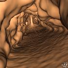
Tracheobronchopathia
osteochondroplastica. CT virtual endoscopy image showing the protruding submucosal nodules of the anterolateral tracheal walls and the preserved airway lumen. The posterior membrane does not present any irregularities.
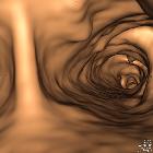
Tracheobronchopathia
osteochondroplastica. CT virtual endoscopy image showing the sporadic protruding submucosal nodules of the anterolateral walls of the right main bronchus
verkalkte tracheale Noduli
Siehe auch:


 Assoziationen und Differentialdiagnosen zu verkalkte tracheale Noduli: