Wolcott-Rallison-Syndrom


Os
odontoideum in wolcott-rallison syndrome: a case series of 4 patients. Radiological images from Case 1. a) Pre-operative sagittal midline CT (on bone windows) shows an os odontoideum widely separated from the body of C2 with a widening of the anterior atlanto-dental distance. There is a marked reduction in the sagittal bony canal dimensions between C2 body and the C1 posterior arch) b) Post-operative sagittal midline CT (on bone windows) demonstrates resection of the posterior arch of atlas and some reduction of the os odontoideum with a reduced distance between the os and the body of C2. c) Lateral plain radiograph demonstrates the occipito-cervical rod and screw fixation

Os
odontoideum in wolcott-rallison syndrome: a case series of 4 patients. Radiological image from Case 2. Midline sagittal T2w image demonstrates lack of ossification of the dens

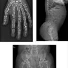
Os
odontoideum in wolcott-rallison syndrome: a case series of 4 patients. Radiological images from Case 3. a Pre-operative CT scan showing C1-2 subluxation with antero-inferior subluxation of C1 lateral mass on C2; (b) pre-operative MRI images showing coronal and sagittal views of C2 demonstrating congenital dens hypoplasia with central dip typical of embryological abnormality and absence of posterior arch of C1; (c) post-operative XR image showing occipital C2 fixation following C1-2 reduction (C1 now in line with C2)
Wolcott-Rallison-Syndrom
Siehe auch:
und weiter:


 Assoziationen und Differentialdiagnosen zu Wolcott-Rallison-Syndrom: