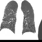
Maximumintensitätsprojektion
Maximum Intensity Projection (MIP) consists of projecting the voxel with the highest attenuation value on every view throughout the volume onto a 2D image .
Such an algorithm is rather simple: for each XY coordinate, only the pixel with the highest Hounsfield number along the Z-axis is represented so that in a single bidimensional image all dense structures in a given volume are observed. For example, it is possible to find all the hyperdense structures in a volume, independently of their position .
This method tends to display bone and contrast material-filled structures preferentially, and other lower-attenuation structures are not well visualized . The primary clinical application of MIP is to improve the detection of pulmonary nodules and assess their profusion. MIP also helps characterize the distribution of small nodules. In addition, MIP sections of variable thickness are excellent for assessing the size and location of vessels, including the pulmonary arteries and veins .
History and etymology
The American radiologist, working in Stanford, California, Sandy Napel (fl. 2019) et al introduced sliding thin-slab maximum intensity projection (STS-MIP) as a new technique in 1993 .
Siehe auch:

 Assoziationen und Differentialdiagnosen zu Maximumintensitätsprojektion:
