aberrant left pulmonary artery


Aberrant left pulmonary artery, also known as pulmonary sling, represents an anatomical variant characterized by the left pulmonary artery arising from the right pulmonary artery and passing above the right main bronchus and in between the trachea and esophagus to reach the left lung. It may lead to compression and focal stenosis of the trachea.
Epidemiology
Associations
Other anomalies that can be associated with aberrant left pulmonary artery are:
- head and neck
- absent thyroid isthmus
- thoracic
- complete tracheal rings
- tracheal stenosis
- single lobed left lung
- bilobed right lung
- congenital lobar overinflation
- abdominopelvic
- musculoskeletal
Clinical presentation
Respiratory distress predominates over esophageal symptoms, usually presenting early in the neonatal period.
Pathology
Pathogenesis
Aberrant left pulmonary arteries are thought to arise from a failure of formation of the 6 aortic arch. They have an anomalous origin from the posterior wall of the right pulmonary artery before coursing to the left lung passing posterior to the trachea and anterior to the esophagus.
The term “sling” is best used when the proximal portion of the anomalous vessel impinges on the right main bronchus and causes air trapping of the entire right lung, or right middle or lower lobes.
The second type of aberrant left pulmonary artery, which often is fatal, is associated with long-segment tracheal stenosis. This kind of tracheal stenosis is due to complete tracheal rings.
Radiographic features
Plain radiograph
Conventional radiographs obtained in neonates at birth may show fetal fluid retention or air, with a mediastinal shift usually to the left side.
In adults, a left-sided deviation of the trachea and an anterior bowing of the right main stem bronchus may be seen. In cases of ring sling complex, radiographs often show an absence of unilateral pulmonary aeration.
Fluoroscopy
In most instances, the barium esophagogram characteristically shows a mass between the trachea and the esophagus just above the level of the carina, usually seen as an anterior indentation over the esophagus.
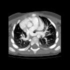
CT/MRI
The main bronchi have horizontal courses (i.e. low T-shaped carina), and vascular anatomy is normally well delineated on CT or MR angiography. Atelectasis may be seen in the upper lobes.
Treatment and prognosis
Repositioning of the artery usually reverses compression, particularly when the underlying tracheobronchial tree is normal.
The mortality rate is high in patients requiring tracheal reconstruction because the stenosis is primary and not due to the vessel.
The success of reconstructive procedures in the rigid trachea can be studied by using three-dimensional CT techniques such as virtual bronchoscopy.
Practical points
- it is the only vascular ring to pass between the trachea and esophagus
- it compresses the trachea posteriorly and causes anterior impression over the esophagus on lateral radiographs
Siehe auch:
und weiter:

