Beckenboden


Dynamic
magnetic resonance imaging of the female pelvic floor—a pictorial review. Normal female pelvic anatomy. Sagittal midline T2-TSE image demonstrating the perineal body (*) and the levator plate (white arrow). B, bladder; U, uterus; V, vagina; R, rectum

Dynamic
magnetic resonance imaging of the female pelvic floor—a pictorial review. Pelvic compartments. Sagittal midline T2-TSE image representing the three pelvic compartments in different colors

Dynamic
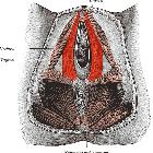
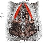
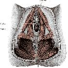
magnetic resonance imaging of the female pelvic floor—a pictorial review. Representative image of the pelvic floor ligaments and their relative anatomical positions in a female model
Beckenboden
Siehe auch:
- Musculus bulbospongiosus
- Musculus ischiocavernosus
- Musculus transversus perinei profundus
- Musculus transversus perinei superficialis
- Beckenbodenmuskulatur
- Musculus coccygeus
- Diaphragma pelvis
- Musculus levator ani
- Musculus sphincter ani externus
- Fossa ischiorectalis
- Schwellkörper- und Schließmuskelschicht
- Diaphragma urogenitale
und weiter:


 Assoziationen und Differentialdiagnosen zu Beckenboden: