Bronchial carcinoid tumor


Bronchial carcinoid tumors are carcinoid tumors primarily occurring in relation to a bronchus. They were previously incorrectly termed as bronchial adenomas. They usually occur in association with a segmental or larger bronchus.
Epidemiology
Typically affects patients from 3 to 7decades with the mean age around 45 years .
Clinical presentation
Presentation can vary dependent on location. Central neoplasms usually give symptoms due to bronchial obstruction (such symptoms can include pneumonia, atelectasis, bronchiectasis, emphysema or even a lung abscess); if airway obstruction is partial, symptoms such as cough, wheezing and recurrent pulmonary infections can occur. Peripheral tumors on the contrary are generally asymptomatic and they are discovered occasionally. Presentation with the carcinoid syndrome is rather rare (~2-5%).
Pathology
They are neuroendocrine neoplasms that range from low-grade typical carcinoids to more aggressive atypical carcinoids. They can therefore demonstrate a wide spectrum in terms of clinical behavior as well as histology.
Location
Most (~60%) tend to be central within the tracheobronchial tree ; the vast majority of which arise from the central bronchi, rarely from the trachea .
Associations
- multiple endocrine neoplasia type 1
- Cushing syndrome: due to ACTH-producing carcinoid tumor types
Radiographic features
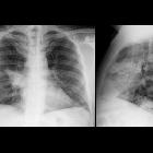
Plain radiograph
Chest radiographic features are often non-specific. They are most frequently seen as round or oval opacities with sharp and often notched margins. Associated airway compression with pulmonary atelectasis may be also seen in some cases.
CT
Central lesions are usually seen as:
- a single hilar or perihilar mass which is usually well-defined, round or ovoid
- can be of any size but are typically 2-5 cm
- there is often marked homogeneous contrast enhancement due to high vascularity
- calcification (usually eccentric) can occur but is not a common feature
Siehe auch:
- Karzinoid
- Neuroendokriner Tumor
- Lungentumor
- bronchiale Tumoren
- peripheral pulmonary carcinoid tumour
- Bronchusobstruktion
- Cushing-Syndrom
- Karzinoid der Trachea
- endobronchial solitary nodule
und weiter:


 Assoziationen und Differentialdiagnosen zu bronchiales Karzinoid: