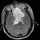
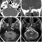
Clival masses


Raumforderungen des Clivus
Clival masses
The differential of a mass involving or arising from the clivus is a relatively narrow one and can be divided into whether the lesion arises from the skull base itself, the intracranial compartment above or the base of skull below.
When evaluating the clivus it is important to compare the marrow signal to that of the adjacent pons on non-contrast, non-fat saturated T1 sequences. Sagittal plane is most useful . See: normal bone marrow signal of the clivus.
Base of skull
- metastasis
- aggressive soft tissue mass
- often patient has a known malignancy
- chordoma
- midline
- bright on T2
- thumb sign
- elevated pituitary out of pituitary fossa
- bony fragments
- ecchordosis physaliphora
- chondrosarcoma
- paramedian, arising from the petro-occipital fissure
- also bright on T2 (chondroid matrix)
- intralesional calcifications in >50%
- elevated pituitary out of pituitary fossa
- plasmacytoma
- T2 signal low to intermediate
- intraosseous lymphoma
- neuroenteric cyst
- can be located with in the base of skull or in the intracranial compartment
- signal intensity varies depending on content
An important normal variant that can sometimes be mistaken for clival pathology is a prominent basilar venous plexus that can appear to erode the posterior surface of the clivus , as well as prominent clival diploic veins that may mimic masses of the body of the clivus .
Intracranial compartment growing into clivus
- invasive pituitary macroadenoma
- arises from pituitary fossa
- no normal pituitary identifiable
- meningioma
- may be intraosseous
- dural tail
- hyperostosis may be present
- craniopharyngioma
Head and neck spaces growing into clivus
- mucocele of the sphenoid sinus
- nasopharyngeal carcinoma
- squamous cell carcinoma of the nasopharynx, sphenoid sinus or posterior ethmoidal air cells
- nasopharyngeal rhabdomyosarcoma
Siehe auch:
- Tornwaldt-Zyste
- Chondrosarkom
- Tumoren der Hypophysenregion
- Chordom
- Makroadenom Hypophyse
- Ecchordosis physaliphora
- Multiples Myelom
- Kraniopharyngeom
- Mucocele
- Dural-Tail-Zeichen
- Nasopharynxkarzinom
- normal bone marrow signal of the clivus
- thumb sign of chordoma
- Osteomyelitis im Clivus
- Meningeom des Klivus
- intrakranielle neuroenterische Zyste
und weiter:


 Assoziationen und Differentialdiagnosen zu Raumforderungen des Clivus:


