congenital cholesteatoma

Congenital
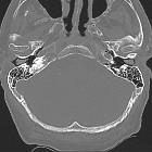
cholesteatoma • Petrous apex cholesteatoma - Ganzer Fall bei Radiopaedia
Congenital
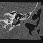
cholesteatoma • Congenital cholesteatoma - Ganzer Fall bei Radiopaedia
Congenital
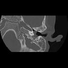
cholesteatoma • Congenital cholesteatoma - Ganzer Fall bei Radiopaedia
Congenital cholesteatomas are identical to epidermoid cysts, differing only in name and location.
Pathology
They are intraosseous inclusions of ectoderm and are therefore comprised of keratin debris and cholesterol. Characteristically, they are located at the petrous apex. In contrast, middle ear cholesteatomas tend to be acquired, secondary to tympanic membrane perforation.
Radiographic features
MRI
Unsurprisingly congenital cholesteatomas have imaging characteristics similar to those of epidermoid cysts
MRI shows sharply demarcated margins with smooth bony erosions.
- T1: low signal
- T2
- high signal (usually slightly brighter than CSF)
- usually will not attenuate on FLAIR imaging, however partial or heterogeneous attenuation may occur. This is in contrast to cholesterol granulomas which never attenuate
- T1 C+ (Gd): no enhancement of the mass itself; thin linear enhancement confined to the margins may be seen
A white epidermoid variant is also recognized in bone, in which case it becomes indistinguishable from a cholesterol granuloma, as both are bright on both T1 and T2 due to the high cholesterol component.
Differential Diagnosis
- chronic otitis media: may be complicated by granulation tissue formation and development of an acquired cholesteatoma
- aberrant internal carotid artery: MRI shows signal flow void, and postcontrast images demonstrate avid arterial enhancement
See also
Siehe auch:
- Cholesterolgranulom der Felsenbeinspitze
- Cholesteatom
- epidermale Inklusionszyste
- Trommelfell
- erworbenes Cholesteatom
- white epidermoid
- middle ear cholesteatomas
und weiter:


 Assoziationen und Differentialdiagnosen zu congenital cholesteatoma:
Cholesterolgranulom
der Felsenbeinspitze