fibromuscular dysplasia


Fibromuscular dysplasia (FMD) is a heterogeneous group of vascular lesions characterized by an idiopathic, non-inflammatory, and non-atherosclerotic angiopathy of small and medium-sized arteries.
Epidemiology
The prevalence is unknown . It is most common in young women with a female to male ratio of 3:1, and is typically diagnosed between the ages of 30 and 50 .
Clinical presentation
Fibromuscular dysplasia is frequently asymptomatic. Symptomatic patients commonly present with:
- hypertension, or less commonly renal impairment, due to renal artery stenosis
- CNS symptoms (e.g. headache, neck pain, pulsatile tinnitus, Horner syndrome) from transient ischemic attack, stroke, dissection, due to carotid and vertebral artery involvement
- angina, myocardial infarction or sudden cardiac death due to coronary artery involvement
- symptoms of mesenteric ischemia (mesenteric infarction is rare due to formation of collateral supply)
Pathology
The exact cause is not well known. The underlying pathology is fibrous or fibromuscular thickening of the arterial wall. Any layer of the vessel wall may be affected: intima, media or adventitia. There is absence of inflammatory cells .
Classification
Fibromuscular dysplasia is classified into five categories according to the vessel wall layer affected:
- intima: 5%
- intimal fibroplasia (see carotid intimal fibromuscular dysplasia)
- media: 90-95%
- medial dysplasia (70%, commonest type)
- perimedial (subadventitial) fibroplasia (15-20%)
- medial hyperplasia (8-10%)
- adventitia: rare
- adventitial fibroplasia (1%)
The outcome is arterial stenoses. Fibromuscular dysplasia most commonly causes small stenoses along a vessel with intervening areas of dilatation (small aneurysms), creating a “string of beads” appearance. Less commonly the stenosis has a smooth tapered appearance. Fibromuscular dysplasia also weakens the vessel wall which predisposes to dissection.
Location
Fibromuscular dysplasia may affect any medium sized artery in the body, and is commonly multifocal and bilateral (up to 60% when involving the renal arteries). Fibromuscular dysplasia usually involves mid segment of the vessels and spares origins. Some sites are more frequently involved :
- renal arteries (most common): estimated between ~5% (range 4-6%) in the renal arteries
- cervicoencephalic arteries (next most common): estimated prevalence ~1.5% (range 0.3-3%)
- extracranial internal carotid arteries
- vertebral arteries
- iliac arteries
- celiac trunk and mesenteric arteries
- subclavian and axillary arteries
Complications
- spontaneous dissection
- distal embolization (of thrombus formed in aneurysm)
- aneurysms
- renal artery aneurysm in ~40% with renal artery fibromuscular dysplasia
- intracranial aneurysms +/- subarachnoid hemorrhage
- arteriovenous fistula
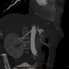
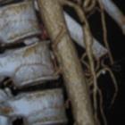
Radiographic features
Arterial imaging with CT angiography, MR angiography and digital subtraction angiography (DSA) may be used to visualize the lesions in fibromuscular dysplasia.
Selective DSA is the gold standard because it allows visualization of small or peripheral lesions. The characteristic finding, particularly in the more common medial subtype, is alternating stenoses and dilatations, causing a string of beads appearance .
Less commonly in intimal and adventitial types, there is focal concentric, long-segment tubular stenosis or diverticular outpouching present (see carotid intimal fibromuscular dysplasia). Cross-sectional imaging (CT and MRI) allows assessment of end-organ ischemic damage.
- typical angiographic features include: vascular loops, fusiform vascular ectasia and a string of beads
- less typical features include: arterial dissection, aneurysm and subarachnoid hemorrhage
Treatment and prognosis
Asymptomatic cases are only observed but if symptomatic then fibromuscular dysplasia responds well to angioplasty, with high long-term patency rates. A stent is generally not required.
Differential diagnosis
Imaging differential considerations include:
- atherosclerosis: usually at origin or proximal portion of the artery
- vasculitides: elevated ESR +/- fever present
- traumatic/iatrogenic vascular injury: correlate with appropriate history
- segmental arterial mediolysis
- vasospasm
Siehe auch:
- Vaskulitis
- Nierenarterienstenose
- Arteria renalis
- Atherosklerose
- renovaskuläre arterielle Hypertonie
- Arteria carotis
- Fibromuskuläre Dysplasie Nierenarterie
- segmentale arterielle Mediolyse (SAM)
- string-of-beads sign
- fibromuscular dysplasia of intracranial arteries
- Fibromuskuläre Dysplasie der Arteria carotis interna
und weiter:
- Aneurysma spurium
- Aneurysma
- Dissektion Arteria vertebralis
- Nierenarterienaneurysma
- Milzarterienaneurysma
- posttraumatisches Aneurysma spurium
- true aneurysm
- stroke in children and young adults
- Aneurysma Arteria iliaca
- vascular pathology
- string of beads sign
- Moyamoya-Muster
- Dissektion Nierenarterie
- Dissektion Truncus coeliacus
- akuter Verschluss Arteria renalis
- Carotis-Sinus-cavernosus-Fistel
- fibromuscular dysplasia classification
- renal balloon angioplasty in fibromuscular dysplasia
- spontane Dissektion Iliakalarterien
- Dissektion der Arteria mesenterica superior
- isolated fibromuscular dysplasia of the middle cerebral artery
- medial fibroplasia
- medial hyperplasia
- intimal fibroplasia
- subadventitial fibroplasia
- intimale Variante der fibromuskulären Dysplasie der Arteria carotis interna
- Fibromuskuläre Dysplasie der Koronarien


 Assoziationen und Differentialdiagnosen zu Fibromuskuläre Dysplasie:


