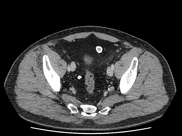
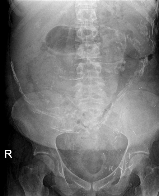
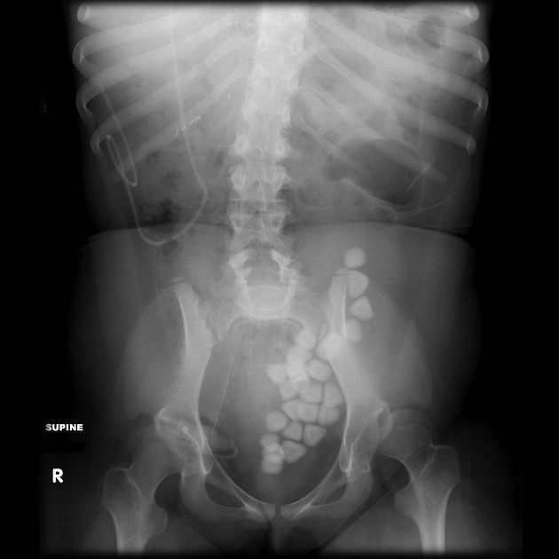
Granulom an chirurgischen Nähten oder Clips
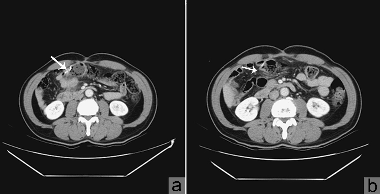
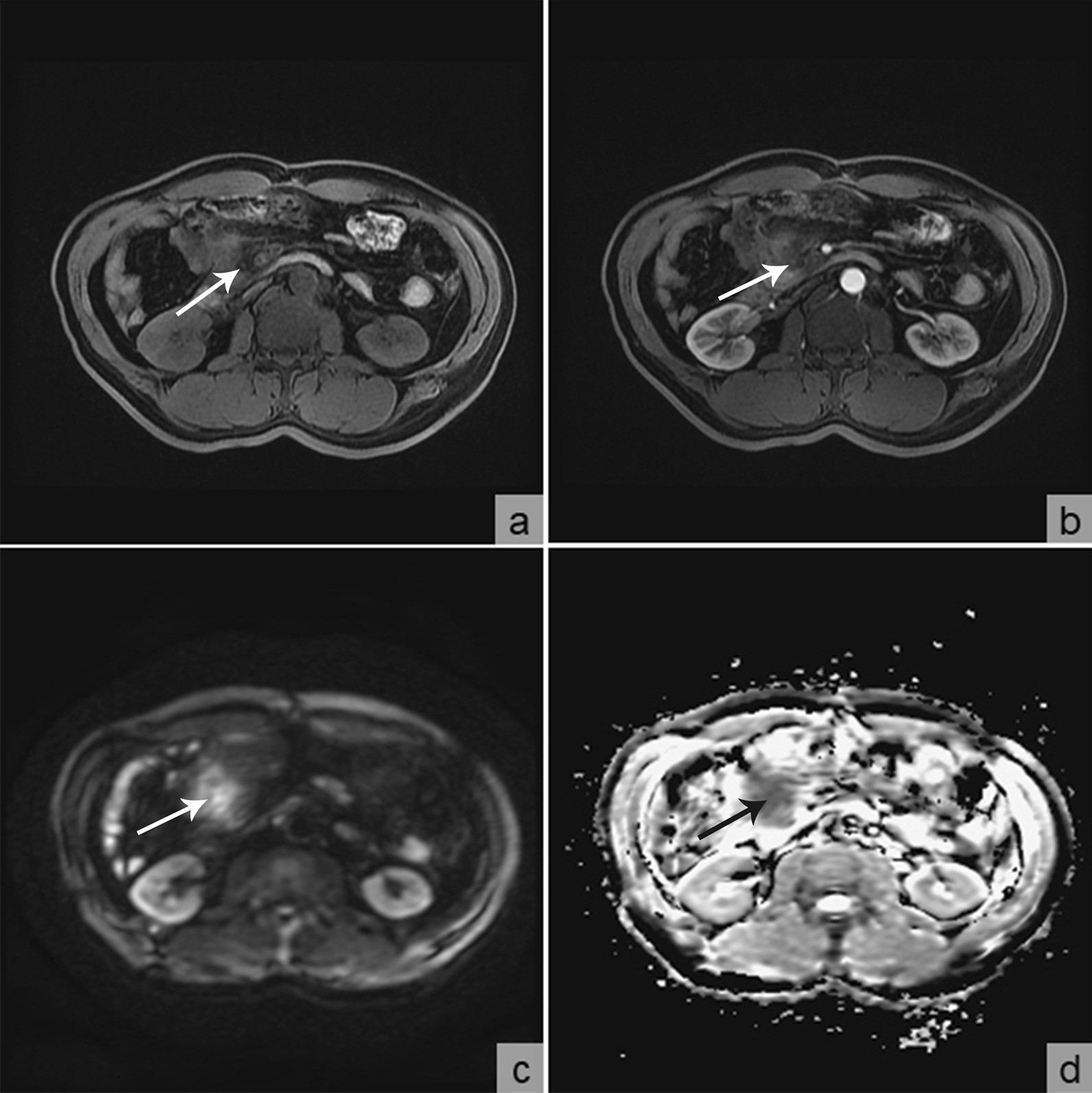
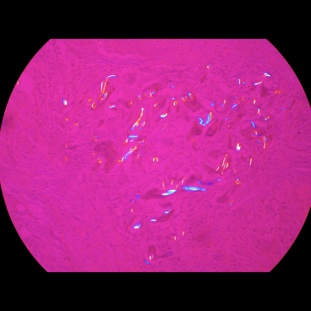
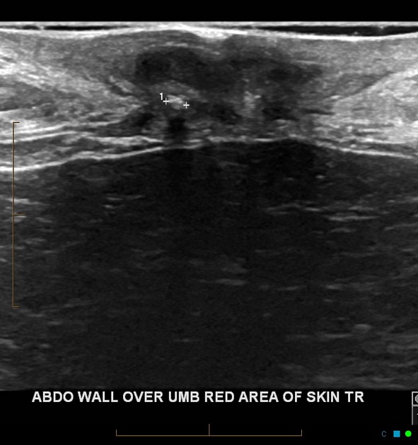
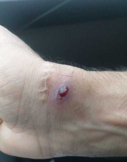
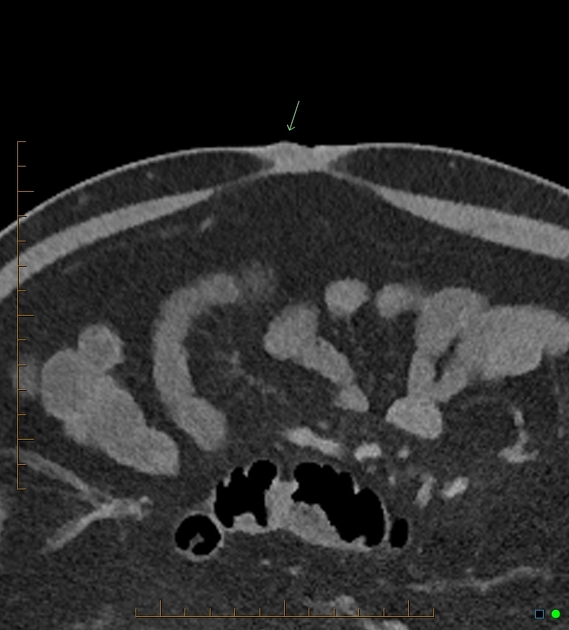
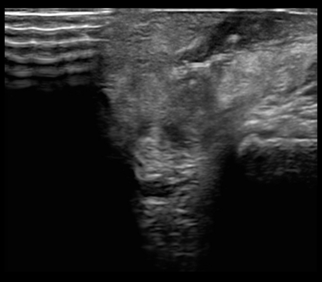
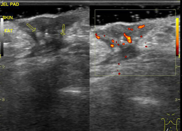
Suture granulomas are localized inflammatory reactions in response to retained suture material. A similar process may also occur in certain situations with mesh repairs . Ultrasound is often used as a first-line imaging modality. It may be confused with a tumor or a recurrent tumor after surgery and should be considered in the differential in the correct setting.
Clinical presentation
Usually develops slowly after an intervention. It may become a palpable and tender mass, mimicking tumor or recurrent tumor.
Pathology
A suture granuloma represents a benign granulomatous proliferation in response to a retained foreign body. They less commonly occur with absorbable sutures, but may still occur.
Radiographic features
Obtaining a history of prior surgery with a surgical approach around the area of concern is important. Suture granulomas can present in the neck after thyroidectomy, mimicking recurrence .
Ultrasound
High-frequency (>10 MHz) linear probe is useful.
- hypoechoic collection
- a small hyperechoic structure in the collection (the suture) is highly specific
- often has parallel hyperechoic 'rail-like' morphology
- may show mild vascularity on color Doppler
PET/CT
- may be FDG avid, mimicking neoplasm
Treatment and prognosis
The treatment of choice is resection of the retained suture and surrounding inflammatory tissue.
Differential diagnosis
General imaging differential considerations include
- recurrent tumor/metastasis
- abdominal desmoid
- scar endometriosis
- abdominal wall abscess
- abdominal wall hematoma
- inflamed epidermoid cyst (internal hair may mimic suture on ultrasound)
Siehe auch:
- verkalkte mesenteriale Lymphknoten
- freie Gewebeformationen peritoneal
- peritoneale Verkalkungen
- intraabdominelle Verkalkungen
und weiter:

 Assoziationen und Differentialdiagnosen zu Granulom an chirurgischen Nähten oder Clips:
