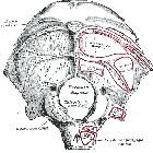
hypoglossal canal


The hypoglossal canal is located between the occipital condyle and jugular tubercle and runs obliquely forwards (posteromedial to anterolateral) allowing the hypoglossal nerve to exit the posterior cranial fossa.
Its proximal portion is often divided by a fibrous (sometimes ossified) septum, which separates the two roots of the hypoglossal nerve (these have formed by the convergence of numerous rootlets). These roots merge within the canal and a single nerve emerges.
Cross-sectional anatomy
The hypoglossal canal is most easily identified on axial or coronal images through the occipital condyles, where it can be seen passing anterolaterally from the posterior fossa into the upper neck.
On coronal imaging, the jugular tubercle appears beak-like extending over the hypoglossal canal. Combined with occipital condyle the coronal appearance is reminiscent of an eagle seen in profile, making this a convenient landmark .
Variant anatomy
- may be enlarged and contain a persistent hypoglossal artery
Siehe auch:
und weiter:


 Assoziationen und Differentialdiagnosen zu Canalis nervi hypoglossi: