Kontrastmittel verstärkte Mammpgraphie


There are 2 types of contrast-enhanced mammography examination – temporal subtraction and dual-energy.
Initial work in the early 2000s used temporal subtraction, but artefacts due to patient movement during prolonged compression limited its diagnostic usefulness. Travieso et al produced a useful comparison table of the 2 techniques in their 2014 paper.
Commercially available contrast mammography equipment now utilizes the dual-energy technique, also known as contrast-enhanced spectral mammography (CESM).
A typical digital mammogram uses energy of ~29kVp. As the K edge of iodine is 33.2 keV, injecting iodinated contrast and performing standard mammography would result in a low signal intensity, indistinguishable from background tissue. CESM is based on dual-energy acquisition – a low energy spectrum, using standard mammography kV and filtration, and a higher energy (above the k-edge of iodine), with stronger filtration.
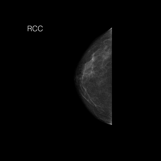
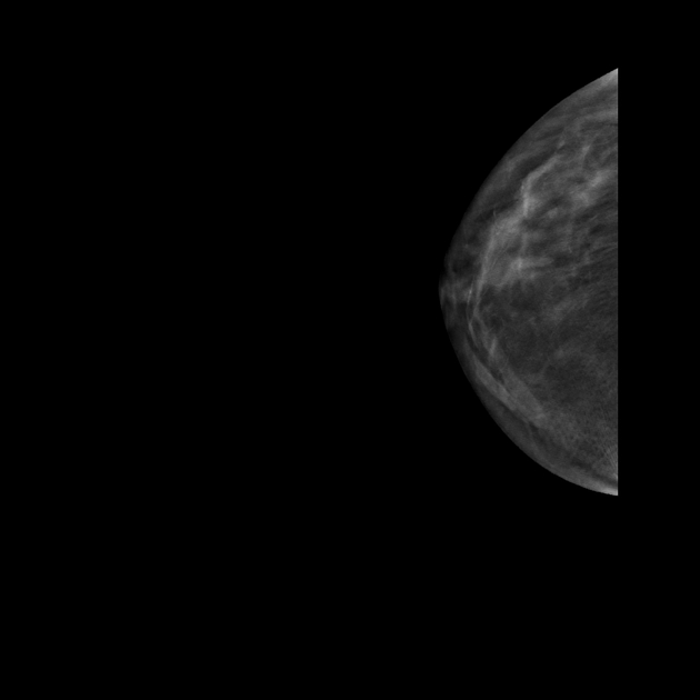
2 images are produced from each compression: low-energy – equivalent to a standard digital mammogram; and recombined - background breast tissue is suppressed to highlight areas of contrast uptake.
The process is straight-forward - iodinated contrast is given (a typical dose is 100 mls of iopamidol 300 at 3mls/second) via a cannula in the antecubital fossa. Approximately 2 minutes later, the first images are acquired. Standard views are obtained – CC and MLO of each breast. The dual-energy exposure occurs during one compression (the exposure is a little longer than a standard examination), so the technique is otherwise identical to an ordinary mammogram.
Previously described as complementary imaging, CESM is now being used instead of a standard digital mammogram in certain situations, e.g. high clinical suspicion of breast cancer. It is also a useful alternative to MRI for problem-solving, or where MRI is contraindicated (e.g. gadolinium allergy or claustrophobia) for staging, or follow-up post-treatment. Research is underway to evaluate its potential use in personalised screening.
Whilst standard digital mammography has a sensitivity of up to 80%, this is lower in dense breasts. CESM has an extremely high sensitivity for invasive breast cancer – up to 98%. Although false-positives do occur (benign lesions can also enhance), the specificity of CESM is generally higher than that of breast MRI.
Siehe auch:

 Assoziationen und Differentialdiagnosen zu Kontrastmittel verstärkte Mammpgraphie:


