Mediastinum
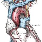
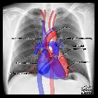

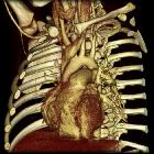
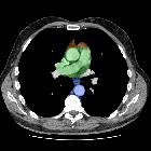

The mediastinum is a space in the thorax that contains a group of organs, vessels, nerves, lymphatics and their surrounding connective tissue. It lies in the midline of the chest between the pleura of each lung and extends from the sternum to the vertebral column.
Gross anatomy
The mediastinum contains all the thoracic viscera except the lungs: heart and great vessels, internal mammary vessels, proximal aspect of azygos venous system, esophagus, trachea, phrenic nerve, cardiac nerve, supra-aortic and para-aortic bodies, thoracic duct, thymus, and mediastinal lymph nodes.
Anatomical division
Division of the mediastinum is generally conceptualized as comprising 3 or 4 compartments, depending on the schema. For example, the mediastinum can be divided into parts based on their relationship to the fibrous pericardium:
- superior mediastinum: above the upper level of the pericardium and plane of Ludwig
- inferior mediastinum: below the plane of Ludwig
- anterior mediastinum: anterior to the pericardium
- middle mediastinum: within the pericardium
- posterior mediastinum: posterior to the pericardium
Historically, there have been discrepant definitions of the mediastinal compartments between medical disciplines. For example, radiologists originally used arbitrary radiologic landmarks on lateral radiographs, while surgeons considered the mediastinum according to anatomic limits encountered intraoperatively (e.g. Shields classification). In response, there have been efforts to reconcile mediastinal classification systems for consistency. The ITMIG classification system, developed by a multidisciplinary group including radiologists and thoracic surgeons, relies on CT-based anatomic divisions.
Relations
- superiorly: continuous with the loose connective tissue of the neck
- anteriorly: chest wall
- laterally: lungs and pleura
- posteriorly: thoracic spine
- inferiorly: diaphragm
Pathology
Broadly, pathology that affects the mediastinum can be divided into those that result in a focal mass, or those that result in diffuse disease involving the mediastinum.
The differential diagnosis of a mediastinal mass is highly dependent on its location within the mediastinum, as this may reveal the structure of origin. Thus, there is a specific differential diagnosis for each compartment:
- thoracic inlet masses
- anterior mediastinal masses
- middle mediastinal masses
- posterior mediastinal masses
In cases where localization is difficult due involvement of multiple compartments, the ITMIG recommends a "center method" technique by which the central point on the axial slice where the lesion appears largest is used for localization. Alternatively, a "structure displacement" technique whereby displacement of organs from adjacent compartments may be used to localize the lesion.
Diffuse mediastinal disease can be separated into groups depending on whether the mediastinal disease is smooth or lobulated:
- smooth
- mediastinal lipomatosis
- mediastinal malignant infiltration
- mediastinal hemorrhage
- mediastinitis
- lobulated
- mediastinal lymphadenopathy
- thymic mass
- germ cell neoplasm
- mediastinal vascular abnormality
- mediastinal neurofibromatosis
Finally, air tracking into the potential mediastinal spaces (pneumomediastinum) may occur when there is rupture of an air-containing structure, penetrating injury, or may be iatrogenic.
Siehe auch:
- Thymus
- Mediastinalemphysem
- Perikard
- Tumoren des vorderen oberen Mediastinums
- mediastinale Raumforderungen
- posterior mediastinal masses
- Lunge
- hinteres Mediastinum
- fetthaltige mediastinale Raumforderungen
- vorderes Mediastinum
- heart and great vessels
und weiter:
- laterale Halszyste
- Arteria carotis communis
- thoracic plane
- danger space
- right paratracheal stripe
- Herz Anatomie
- posterior mediastinal mass in the exam
- Thorax Onlinekurs
- Zyste oder Fistel des vierten Kiemenbogens
- oberes Mediastinum
- Zyste oder Fistel des ersten Kiemenbogens
- mittleres Mediastinum
- Mediastinalschatten


 Assoziationen und Differentialdiagnosen zu Mediastinum:


