Meyersche Dysplasie

Preschooler
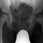
with left hip pain. AP radiograph of the hips shows a normal appearing left hip. There is flattening and sclerosis of the right femoral epiphysis which may have multiple ossification centers and there is also right metaphyseal subcortical cystic change.The diagnosis was Meyer dysplasia.
Meyer dysplasia (also known as dysplasia epiphysealis capitis femoris) is a fragmentation and delayed ossification of the femoral capital epiphyses that affects the pediatric hips. It is considered more of a normal hip developmental variation rather than a true dysplasia. It can be bilateral in ~50% of cases .
Epidemiology
It tends to present at around 2-3 years of age . There is a recognized male predilection .
Clinical presentation
Patients are usually asymptomatic.
Pathology
There is a delay in development of the nucleus of ossification in the hip which does not occur until ~1.5 years of age. The epiphysis does not collapse and density and structure remain preserved. It usually resolves spontaneously later in childhood.
Radiographic features
Plain radiograph
- the affected epiphysis is smaller in size
- there are often multiple nuclei of ossification, giving the epiphysis a "morulated" appearance; these then tend to fuse at ~5 years of age.
History and etymology
First described by J Meyer in 1964 .
Differential diagnosis
On imaging consider
Siehe auch:


 Assoziationen und Differentialdiagnosen zu Meyersche Dysplasie: