Neonatal pneumothorax


Neonatal pneumothorax describes pneumothoraces occurring in neonates. It is a life-threatening condition, associated with high morbidity and mortality. The diagnosis is a challenge especially when the amount of air is small and may accumulate along the anterior or medial pleural space.
Epidemiology
Only 0.5% of cases are symptomatic despite the high incidence. Studies show a male predilection. It is more commonly seen to occur within the first three days of life with more than 80% seen in the first 48 hours .
Clinical presentation
Variable from being completely asymptomatic to severe respiratory distress. Symptoms include dyspnea, cyanosis and chest pain. Physical examination may reveal decreased breath sounds, hyper-resonance to percussion on the afflicted side with contralateral tracheal deviation. When under tension, even cardiac function may be compromised.
Pathology
Air dissects from alveolar spaces into interstitial spaces of lung. The eventual migration of air to the visceral pleura culminates in rupture into the pleural cavity. It may also be accompanied by pneumomediastinum, pneumopericardium, pneumoperitoneum or subcutaneous emphysema.
Etiology
It may occur spontaneously, however, more commonly seen secondary to birth trauma, positive pressure ventilation, underlying obstructive or restrictive lung disease and rupture of a congenital or acquired cyst .
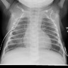
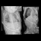
Radiographic features
Two signs are useful in the diagnosis of a small pneumothorax
- hyperlucent hemithorax sign in case of anterior pneumothorax
- medial stripe sign in case of medial pneumothorax
Views
These signs can be appreciated in the supine position with additional views to confirm:
- hyperlucent hemithorax sign is seen as unilateral or bilateral areas of increased lucency confirmed on cross-table lateral view; the air will be seen to accumulate anteriorly
- medial stripe sign is seen as an area of increased lucency at the interface of the medial lung and the mediastinum which can be confirmed on lateral decubitus view; the air will be seen to rise and accumulate along the lateral chest wall
Treatment and prognosis
Small, asymptomatic pneumothoraces are left alone and kept under close observation. Larger or symptomatic ones require drainage. Acute tension may be relieved with needle aspiration; chest tube or pigtail drainage may be placed afterwards for complete evacuation.
See also
Siehe auch:


 Assoziationen und Differentialdiagnosen zu Pneumothorax beim Neugeborenen: