pineal gland


 nicht verwechseln mit: Epiphyse des Knochens
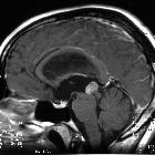
The pineal gland is a small, pine-cone shaped structure considered to be part of the epithalamus. It is unpaired and situated in the midline.
Gross anatomy
The pineal gland typically measures around 7 x 6 x 3 mm in size and is situated in a groove between the laterally placed thalamic bodies . Normal sizes have been described in the radiology literature up to 14 mm . Appearing to arise from the gland are two laminae. Above is the habenular commissure and below it is the posterior commissure.
Relations
- anteriorly: third ventricle (pineal recess)
- anterosuperiorly: habenular
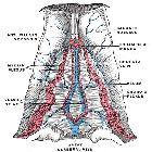
- superiorly: internal cerebral veins, vein of Galen (posteriorly), stria medullaris, splenium of the corpus callosum and velum interpositum
- posteroinferiorly: superior cerebellar cistern
- inferiorly: superior colliculi of the midbrain
- anteroinferiorly: posterior commissure
Blood supply
The pineal gland receives its blood supply from fine branches of the posterior choroidal arteries and drains superiorly by multiple branches eventually into the great cerebral vein of Galen .
Innervation
Sympathetic: superior cervical ganglion
Parasympathetic: otic and pterygopalatine ganglion
Also has central innervation directly through the pineal stalk and also from the trigeminal ganglion which contains the neuropeptide PACAP (pituitary adenylate cyclase-activating polypeptide).
Embryology
The pineal gland arises during the seventh week of gestation from a thickening of the ependyma at the posterior-most aspect of the third ventricle .
Physiology
The pineal gland produces melatonin which affects the modulation of wake/sleep patterns and photoperiodic (seasonal) functions. It is also thought to have a reproductive function and has been associated with the onset of puberty . Unlike much of the rest of the brain, it is not isolated from the body by the blood-brain barrier.
Radiographic features
Plain radiograph
The pineal gland has a predilection for calcification which is invariably histologically present in adults but rarely seen below the age of 10 years . Calcification is visible on lateral skull x-rays in 50-70% of adults . The habenular commissure also calcifies and is visible as a small C-shaped (open part posteriorly) density above and in front of the pineal calcification.
CT
The far higher contrast resolution of CT means that calcification is almost always visible in the adult pineal gland, sometimes visible as specks embedded in a small soft tissue nodule similar in density to grey matter. In children under the age of 5 years, no calcification is present but prevalence increases rapidly with age, reaching a plateau at about 30 years of age .
MRI
MRI is the modality of choice for evaluating the pineal region. Although sensitivity to calcification on conventional sequences is poor, susceptibility weighted imaging (SWI) has similar diagnostic power to CT.
The gland appears as a small nodule of tissue with similar intensity to grey matter. As it lies outside the blood-brain barrier, it demonstrates vivid contrast enhancement.
Related pathology
Siehe auch:
- verkalktes Corpus pineale
- Pinealiszyste
- Corpus callosum
- Tumoren der Pinealisregion
- velum interpositum
- Pineoblastom
- Pineozytom
- intrakraniale Teratome
- Vena cerebri magna
- Epithalamus
- posterior choroidal artery
- Parenchymtumor der Glandula pinealis mit intermediärer Differenzierung
- intrakranielles Germinom
- Suszeptibilitätsgewichtete Bildgebung
- habenula
- Papillärer Tumor der Pinealisregion
- normale intrakranielle Verkalkungen
- Bluthirnschranke
- primary intracranial choriocarcinoma
- Parenchymtumoren der Glandula pinealis
- intrakranieller Keimzelltumor
- intrakranielles embryonales Karzinom
- thalamic
und weiter:


 Assoziationen und Differentialdiagnosen zu Glandula pinealis: