superior ophthalmic vein
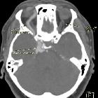
Caroticocavernous
fistula • Caroticocavernous fistula - Ganzer Fall bei Radiopaedia

Superior
ophthalmic vein • Orbital apex (diagram) - Ganzer Fall bei Radiopaedia

Superior
ophthalmic vein • Orbital veins (Gray's illustration) - Ganzer Fall bei Radiopaedia

Superior
ophthalmic vein • Superior ophthalmic vein thrombosis - Ganzer Fall bei Radiopaedia

Superior
ophthalmic vein • Juvenile nasopharygeal angiofibroma - Ganzer Fall bei Radiopaedia
The superior ophthalmic vein is a prominent vein of the orbit that is seen on CT and may be enlarged or tortuous in various disease entities.
Gross anatomy
Origin
Formed in the anterior part of the orbit by the union of the angular, supraorbital and supratrochlear veins.
Course
- courses laterally within the intraconal space (with the ophthalmic artery) to travel between the superior rectus muscle above and the optic nerve and ophthalmic artery below
- exits the intraconal space to become extraconal
- exits the orbit via the superior orbital fissure superior to the annulus of Zinn between the frontal (branch of V1) and trochlear nerves
Termination
- drains directly into the cavernous sinus
Tributaries
- vortex veins draining choroid
- central retinal vein
- veins that correspond to branches of the ophthalmic artery (ethmoidal vein, lacrimal)
Drains
- globe, upper eyelid, extraocular muscles and lacrimal gland
The mean diameter of the vein is 2 mm and normal sizes range from 1 to 2.9 mm . A threshold value of 2.5 mm has also been proposed .
Related pathology
Enlarged or tortuous superior ophthalmic veins may be seen in:
- carotid-cavernous fistula
- ophthalmic vein varix
- raised intracranial pressure
- Graves disease
- orbital pseudotumor
- superior ophthalmic vein thrombosis
Reversible bilateral enlargement of the superior ophthalmic veins is often seen in intubated patients . The superior ophthalmic veins may be hyperdense on CT due to thrombosis, including in cases of orbital cellulitis.
Siehe auch:
- Sinus cavernosus
- Carotis-Sinus-cavernosus-Fistel
- dilatierte Vena ophthalmica superior
- Thrombose der Vena ophthalmica superior
und weiter:


 Assoziationen und Differentialdiagnosen zu Vena ophthalmica superior:
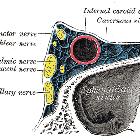

dilatierte
Vena ophthalmica superior
