synoviale Chondromatose Knie

Synoviale
Chondromatose mit multiplen Chondromen in einer Bakerzyste bei Gonarthrose

Synovialchondrome
in Bakerzyste bei Gonarthrose. Mit im Bild eine Kalibrierungskugel für die Vorbereitung zur Prothesenimplantation.


Synovialchondromatose
Kniegelenk: dorsomedial, somit am ehesten in einer Baker-Zyste.

Synovialchondromatose
in einer Baker-Zyste links im Röntgenbild, rechts beginnend einige Jahre vorher in der Magnetresonanztomografie.
Hoffa’s fat
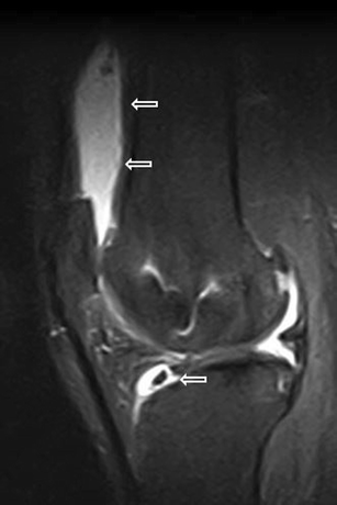
pad abnormalities, knee pain and magnetic resonance imaging in daily practice. Synovial chondromatosis. Sagittal proton density with fat saturation image shows joint effusion with loose bodies in the suprapatellar pouch and in the infra-hoffatic recess (wide arrows). The HFP is oedematous

Synovialchondromatose
Kniegelenk. Zusätzlich ausgeprägte lateral betonte, deformierende Gonarthrose. Weiterhin auch Retropatellararthrose und Stieda-Pellegrini-Schatten.
synoviale Chondromatose Knie
Siehe auch:


 Assoziationen und Differentialdiagnosen zu synoviale Chondromatose Knie: