total anomalous pulmonary venous return


Total anomalous pulmonary venous return (TAPVR) is a cyanotic congenital heart anomaly with abnormal drainage anatomy of the entire pulmonary venous system. This contrasts with partial anomalous pulmonary venous return (PAPVR) where only part of the pulmonary venous anatomy is abnormal.
In TAPVR, all systemic and pulmonary venous blood enters the right atrium and nothing drains into the left atrium. A right-to-left shunt is required for survival and is usually via a large patent foramen ovale (PFO) or less commonly atrial septal defect (ASD).
Clinical presentation
Affected infants develop cyanosis and congestive heart failure in the early neonatal period.
Pathology
The anomaly occurs secondary to an embryological failure of pulmonary venous development:
- at the earliest stage of lung development, the primitive lung bud venous drainage occurs via the splanchnic plexus to primitive systemic (cardinal/umbilicovitelline) veins; at this point, the heart is still forming and there is no pulmonary connection
- at 27 to 29 days of life, a common pulmonary vein begins growing outwards from the primitive left atrium to join the primitive pulmonary veins
- the common pulmonary vein joins with the pulmonary veins from each lung, forming the familiar configuration; simultaneously, there is involution of the primitive connection between pulmonary veins and primitive systemic veins
Failure to completely transition between primitive and mature configurations according to this sequence results in persistent venous drainage from the lungs to one of several possible systemic veins (see Classification) .
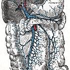
Classification
TAPVR can be classified into four types (in decreasing order of frequency) depending on the site of anomalous venous union :
Type I: supracardiac
- most common type (over 50% of cases)
- anomalous pulmonary veins terminate at the supracardiac level
- pulmonary veins converge to form a left vertical vein which then drains to either brachiocephalic vein, SVC, or azygous vein
Type II: cardiac
- second most common (~30% of cases)
- pulmonary venous connection at the cardiac level
- drainage is into the coronary sinus and then the right atrium
Type III: infracardiac
- connection at the infracardiac level
- the pulmonary veins join behind the left atrium to form a common vertical descending vein
- the common descending vein courses anterior to the esophagus passes through the diaphragm at the esophageal hiatus and then usually join the portal system
- drainage is usually into the ductus venosus, hepatic veins, portal vein, or IVC
Type IV: mixed pattern
- least common type
- anomalous venous connections at two or more levels
Associations
Approximately one-third of those with TAPVR also have other associated cardiac lesions; many have heterotaxy syndrome, particularly asplenia. Type III (infracardiac) is also associated with thoracic lymphangiectasia and pulmonary congestion.
Radiographic features
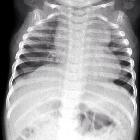
Plain radiograph
The right heart is prominent in TAPVR because of the increased flow volume, but the left atrium remains normal in size. Types I and II result in cardiomegaly.
The supra cardiac variant (type I) can classically depict a snowman appearance on a frontal chest radiograph, also known as figure of 8 heart or cottage loaf heart . The dilated vertical vein on the left, brachiocephalic vein on top, and the superior vena cava on the right form the head of the snowman; the body of the snowman is formed by the enlarged right atrium.
Echocardiography
May show blind-ended left atrium with no connecting veins.
CT/MRA
Direct visualization of anomalous venous return.
Siehe auch:
- Lungensequester
- Lungenvenenfehlmündung
- Atriumseptumdefekt
- Vena portae
- Vena cava inferior
- Lungenhypoplasie
- Scimitar-Syndrom
- Situs ambiguus
- Snowman heart
- Vena cava superior
- partielle Lungenvenenfehlmündung
- Lebervenen
- pulmonary venolobar syndrome
- Zyanotischer Herzfehler
und weiter:


 Assoziationen und Differentialdiagnosen zu totale Lungenvenenfehlmündung: