trochlear nerve


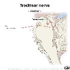


The trochlear nerve is the fourth cranial nerve and is the motor nerve of the superior oblique muscle of the eye. It can be divided into four parts:
Gross anatomy
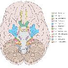
Nucleus and intraparenchymal portion
The trochlear nucleus is located in the dorsoventral midbrain, ventral to the periaqueductal grey matter. Its fibers course dorsally and decussate dorsal to the periaqueductal grey matter before exiting the brainstem immediately below the inferior colliculus. It is the only cranial nerve to exit the brainstem posteriorly.
Cisternal portion
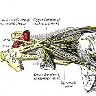
The nerve rounds the cerebral peduncles in the ambient cistern. Eventually, along with the oculomotor nerve (CN III), it courses anteriorly between the superior cerebellar artery below and posterior cerebral artery above before piercing the dura between the free and attached edge of tentorium cerebelli.
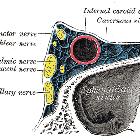
Cavernous sinus portion
Within the cavernous sinus, the trochlear nerve is located initially below the oculomotor nerve in the lateral wall of the sinus, although by the time it reaches the superior orbital fissure, it lies above it (outside the tendinous ring). It is the "Tarts" in this infamous mnemonic.
Orbital portion
It enters the orbit outside the tendinous ring, between the superior ophthalmic vein and the superolateral quadrant of the ring. Once in the orbit, it arches up and medially above superior rectus and levator palpebrae superioris to innervate the superior oblique.
Radiographic features
MRI
Although the nerve is very thin, on high-resolution T2 weighted imaging (e.g. FIESTA/CISS) it can be visualized as a very thin structure. Dedicated higher-resolution sequences can also be performed if greater detail is required .
Related pathology
Siehe auch:
- Fissura orbitalis superior
- Sinus cavernosus
- Hirnnerven
- Anatomie Hirnstamm
- Arteria cerebelli superior
- ambient cistern
- cerebral peduncles
- Annulus of Zinn
- Musculus obliquus superior
- Orbita
- Musculus rectus superior
und weiter:


 Assoziationen und Differentialdiagnosen zu Nervus trochlearis: