uterine tubes


 nicht verwechseln mit: Fallopio-Kanal
The Fallopian tube, also known as the uterine tube or less commonly the oviduct, is a paired hollow tube that bridges between each ovary and the uterus and functions to convey the mature ovum from the former to the latter. If conception occurs, it normally does so within the tube. It can be affected by a wide range of pathology.
Gross anatomy
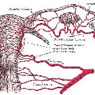
The Fallopian tube is approximately 10-12 cm long and 1-4 mm in diameter. It bridges the gap between the ovary laterally, and the uterus medially. Through it, the ovum passes into the uterine cavity. The peritoneal reflection draping over the salpinges forms the mesosalpinx.
The Fallopian tube is divided into 5 anatomic segments (from lateral to medial, through the path of an ovum after ovulation):
- fimbriae: ~25 finger-like projections that drape over the ovary
- the ovarian fimbriae are longer than the others and are attached to the tubal pole of the ovary
- infundibulum: a funnel-shaped lateral part that drapes over the ovary with the fimbriae emanating from it
- it opens into the peritoneal cavity at the abdominal ostium
- ampulla: the widest and longest section, forming over half the length
- isthmus: immediately lateral to the uterus, it is the narrowest segment, as its name suggests
- interstitial or intramural segment: the section within the myometrium
See mnemonic here.
Relations
The extrauterine part of the Fallopian tube courses between the two folds of the broad ligament, at its superior aspect .
- at its medial end, the interstitial segment is continuous with the uterotubal junction, a continuation of the endometrial cavity
- at its lateral end, the infundibulum opens into the peritoneal cavity
Blood supply
- arterial supply: tubal branch of the ovarian artery (lateral one-third) and terminal (tubal) branch of the uterine artery (medial two-thirds)
- venous drainage:
- lateral one-third via the pampiniform plexus to the ovarian veins
- medial two-thirds via the uterine plexus to the internal iliac vein
Lymphatic drainage
- via ovarian vessels to the para-aortic nodes and uterine vessels to the internal iliac chain
- some drainage also to the inguinal nodes via the round ligament
Innervation
- autonomic supply from the ovarian and uterine plexuses
- parasympathetic: vagus for the lateral half, pelvic splanchnic to the medial half
- sympathetic from the thoracic and lumbar spinal segments (T10- L1)
Variant anatomy
- absent unilateral
- hypoplastic unilateral
- hypoplastic bilateral
- abnormal entry into uterine body or fundus
Histology
Like many other muscular hollow tubes, it has two layers of muscle (inner circular, outer longitudinal), and is lined by a mixture of ciliated and non-ciliated columnar epithelium. It is the former that pushes the ovum towards the uterus.
Radiographic features
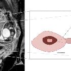
The normal Fallopian tube is not visualized at cross-sectional imaging unless it is outlined by fluid. In the presence of peritoneal fluid or contrast material, the Fallopian tubes appear as paired, thin, serpentine juxta-uterine structures extending either anteriorly or posteriorly into the cul-de-sac.
Fluoroscopy
Contrast studies can be completed by performing a hysterosalpingogram (HSG).
History and etymology
The fallopian tube is named after Gabriel Fallopius, an Italian anatomist (1523-62). He is the same anatomist who gave his name to the Fallopian ligament and the Fallopian canal. Despite the eponym, the word "fallopian" is most often used in lower case.
Related pathology
- tubal carcinoma
- tubal rupture
- tubal torsion
- tubal ectopic pregnancy
- tubo-ovarian abscess
- salpingitis
- hematosalpinx
- hydrosalpinx
- pyosalpinx
Siehe auch:
- Uterus
- tubal ectopic pregnancy
- Tuboovarialabszess
- Hysterosalpingographie
- salpingitis isthmica nodosa
- ovarian artery
- Ovar
- Tubentorsion
- Adnexitis
- Tubenkarzinom
- Tubenruptur
- Arteria uterina
- weibliche Fortpflanzungsorgane
- fallopian tube recanalization
und weiter:
- Pyosalpinx
- adnexal ectopic pregnancy
- Becken
- peritoneal cavity
- knöchernes Becken
- pelvic peritoneal space
- cornual ectopic pregnancy
- ovarian ectopic pregnancy
- Müller-Gang
- angular ectopic pregnancy
- fallopian tube polyp
- gynäkologisch radiologisches Curriculum
- cobblestoning
- haematosalpinx
- fallopian tube selective catheterization and disobstruction


 Assoziationen und Differentialdiagnosen zu Eileiter: