Tumoren der Mamille

Multimodality
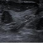
approach to the nipple-areolar complex: a pictorial review and diagnostic algorithm. Syringomatous tumor of the nipple. A 47-year-old woman with retraction and hardening of the left nipple. a Mammogram shows retraction of the left nipple and asymmetric retroareolar density. b On ultrasound, the lesion is solid and hypoechogenic with ill-defined borders and increased peripheral color-Doppler signal. c Contrast-enhanced coronal T1-weighted MRI (subtracted image obtained 120 s after contrast injection) shows mass-type uptake with pronounced early enhancement in a lesion with hazy borders that retracts the nipple-areolar complex. d Hematoxylin-eosin stain (× 4) shows a proliferation of elongated glandular structures like strings of cells in the dermis, with the formation of keratin cysts (star)

Multimodality
approach to the nipple-areolar complex: a pictorial review and diagnostic algorithm. Paget’s disease (I). A 56-year-old woman with right nipple retraction. a 2D mammogram shows a spiculated retroareolar mass in the right breast with nipple retraction and skin thickening; b on ultrasound, it is seen as a solid lesion with ill-defined borders. c MRI shows the retroareolar lesion extending to the nipple-areolar complex. Histologic study revealed infiltrating ductal carcinoma extending to the epidermis

Multimodality
approach to the nipple-areolar complex: a pictorial review and diagnostic algorithm. Nipple adenoma. An 81-year-old woman with a several-week history of pain and swelling of the right breast. a Mammogram shows an isodense rounded retroareolar mass with slightly irregular margins (arrows); b on ultrasound, it is seen as a solid nodular lesion (N: nipple). c Immunohistochemistry stain with p63 (× 4) shows glandular and ductal proliferation consisting of epithelial and myoepithelial cells, which express p63
Tumoren der Mamille
Siehe auch:
und weiter:

 Assoziationen und Differentialdiagnosen zu Tumoren der Mamille:

