Fehlfunktion Fehllage Komplikationen peripher gelegter ZVK (PICC)

Infant after
left lower extremity PICC removal which after measuring was found to be too short in length. AXR AP (left) and lateral (right) show a discontinuous catheter fragment in the left femoral and iliac veins.The diagnosis was peripherally inserted central venous catheter malfunction with the catheter having broken during removal with a retained catheter fragment in the left femoral and iliac veins.

Premature
newborn with a new PICC line. CXR shows a left upper extremity catheter which courses over the left side of the spine and whose tip projects at T12.The diagnosis was peripherally inserted central catheter malposition with the tip in the descending aorta.

Newborn
status post left upper extremity PICC placement. CXR AP shows an umbilical arterial catheter in the aorta whose tip is at the level of the T8 vertebral body and a left upper extremity PICC which courses parallel to the umbilical arterial catheter and whose tip projects at the level of T8 as well.The diagnosis was peripherally inserted central catheter malfunction with the tip of the catheter in the descending aorta.

Newborn after
PICC placement. AXR AP (left) shows the tip of a right lower extremity PICC line projecting at the level of the T11 vertebral body. AXR lateral (right) shows the tip of the PICC line projecting over the spinal canal. The patient also has a large anterior abdominal wall defect containing loops of bowel which is covered by a membranous sac.The diagnosis was peripherally inserted central venous catheter malfunction with the tip within the spinal canal in a patient with an ompahlocele.

Infant who
has just had a PICC line placed. AXR shows a left lower extremity PICC line whose tip projects in the region of the right renal vein.The diagnosis was peripherally inserted central venous catheter malfunction with the tip of the PICC line being in the right renal vein.

Premature
newborn after scalp PICC placementCXR AP shows a right scalp PICC which crosses the midline and whose tip projects over the left pedicle of the spine at T7 either in the aorta or in the hemiazygous vein.The diagnosis was malposition of the PICC, which is either within the aorta or the hemiazygous vein.
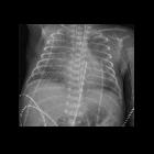
Premature
newborn after PICC placementCXR AP shows an umbilical arterial catheter in the aorta whose tip is at T6 and a left upper extremity PICC which courses parallel to the umbilical arterial catheter and whose tip projects at the level of L2.The diagnosis was malposition of the PICC within the descending aorta.
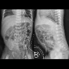
Premature
newborn after PICC placementAXR AP shows a right lower extremity PICC whose tip projects at the level of L1 and that projects within the spinal canal on the lateral radiograph.The diagnosis was malposition of the PICC within the spinal canal.
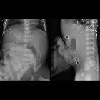
Premature
newborn after PICC placementAXR AP shows a left lower extremity PICC that does not follow the course of the iliac vein and whose tip projects at the level of L3 and that projects within the spinal canal on the lateral radiograph. The only gas within the abdomen is in the stomach while all of the small bowel is in a silo outside the abdomen.The diagnosis was malposition of the PICC within the spinal canal in a patient with gastroschisis.

Preschooler
after PICC placementCXR AP and lateral shows a left upper extremity PICC whose tip projects over the left internal mammary vein.The diagnosis was malposition of the PICC.
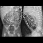
Premature
newborn after PICC placementAXR AP and lateral shows a left lower extremity PICC coursing along in a thoracoepigastric vein along the left lateral abdominal wall. The umbilical arterial catheter tip projects at T10.The diagnosis was malposition of the PICC and appropriate position of the umbilical arterial catheter.
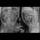
Premature
newborn after PICC placementAXR AP and lateral shows a right lower extremity PICC tip projecting over a superficial epigastric vein in the anterior abdominal wall.The diagnosis was malposition of the PICC.

Premature
newborn after PICC line placement. AXR AP shows a right lower extremity PICC line whose tip projects over the scrotum, presumably in the left gonadal vein.The diagnosis was peripherally inserted central catheter malposition with the tip in the left gonadal vein.

Premature
newborn after PICC line placement. CXR AP shows a left upper extremity PICC line whose tip projects over the left pulmonary artery.The diagnosis was peripherally inserted central catheter malposition in the left pulmonary artery.

Infant with
an omphalocele after lower extremity PICC line placement. AXR AP (left) shows a wavy appearance to the right lower extremity PICC line. Lateral radiograph (right) shows the PICC line tip projecting over the spinal canal.The diagnosis was peripherally inserted central catheter malfunction with the tip of the PICC line in a spinal vein.
Fehlfunktion Fehllage Komplikationen peripher gelegter ZVK (PICC)
Siehe auch:

 Assoziationen und Differentialdiagnosen zu Fehlfunktion Fehllage Komplikationen peripher gelegter ZVK (PICC):


