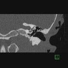
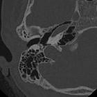
chronische Otomastoiditis mit Tympanosklerose

Myringosclerosis
• Myringosclerosis - Ganzer Fall bei Radiopaedia

External and
middle ear diseases: radiological diagnosis based on clinical signs and symptoms. Chronic middle ear inflammation. Axial CT in bone window demonstrates focal calcifications (arrow) in the tympanic cavity, close to ossicular chain. This is called tympanosclerosis
Chronic otomastoiditis with tympanosclerosis represents calcific foci within the middle ear or tympanic membrane secondary to suppurative chronic otomastoiditis.
Radiographic features
Features include chronic otomastoidits findings such as middle ear soft tissue density and underpneumatised mastoid associated with calcifications. Common locations of calcifications include:
- tympanic membrane
- ossicle surface
- stapes footplate
- muscle tendons
- ossicle ligaments
See also
Siehe auch:
und weiter:

 Assoziationen und Differentialdiagnosen zu chronische Otomastoiditis mit Tympanosklerose:


