Stapes

Middle ear
• Inner ear (illustrations) - Ganzer Fall bei Radiopaedia


Stapes •
Stapes (illustration) - Ganzer Fall bei Radiopaedia

Stapes •
Oval window and stapes - Ganzer Fall bei Radiopaedia

Tympanic
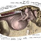
membrane • Middle ear anatomy: diagrams - Ganzer Fall bei Radiopaedia

External and
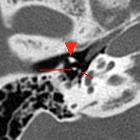
middle ear diseases: radiological diagnosis based on clinical signs and symptoms. Normal anatomy of the middle ear. Axial CT, bone window. Two dots, visible posteriorly to malleus neck (arrowhead) represent incus lenticular process (long arrow) and stapes head (short arrow)

Middle ear
ossicles • Stapes: annotated CT - Ganzer Fall bei Radiopaedia
The stapes (plural: stapedes) is the smallest and most medial of the three middle ear ossicles. It is the smallest bone in the standard human skeleton.
It has a base (foot piece/footplate) which articulates with the oval window and conducts vibrations to the cochlea. The base is attached to the neck via anterior and posterior arches (also called the anterior and posterior crura). On top of the neck (or hub) is a concave head (also known as the capitulum) which articulates with the lentiform nodule (lenticular process) of the incus. The combination of the capitulum, neck, and crura is collectively referred to as the stapes superstructure.
The stapes has a number of attachments:
- incudostapedial joint (synovial joint)
- annular ligament around the oval window, forming the stapediovestibular joint
- stapedius (muscle attaches to the back of the neck of the stapes)
History and etymology
"Stapes" is Latin for stirrup.
Siehe auch:
und weiter:

 Assoziationen und Differentialdiagnosen zu Stapes:


