Incus

Middle ear
• Inner ear (illustrations) - Ganzer Fall bei Radiopaedia


Incus •
Incus: anatomy illustration - Ganzer Fall bei Radiopaedia

Incus •
Incus: annotated CT - Ganzer Fall bei Radiopaedia

Tympanic
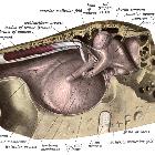
membrane • Middle ear anatomy: diagrams - Ganzer Fall bei Radiopaedia

External and
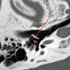
middle ear diseases: radiological diagnosis based on clinical signs and symptoms. Normal anatomy of middle ear. Axial CT, bone window. Malleus head (long arrow) and incus body (short arrow) with short process are visible in epitympanum, forming a so-called “ice cream cone”

External and
middle ear diseases: radiological diagnosis based on clinical signs and symptoms. Normal anatomy of middle ear. Axial CT, bone window. Malleus handle (long arrow) and incus long process (short arrow) are visible, forming two parallel lines

External and
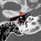
middle ear diseases: radiological diagnosis based on clinical signs and symptoms. Normal anatomy of the middle ear. Axial CT, bone window. Two dots, visible posteriorly to malleus neck (arrowhead) represent incus lenticular process (long arrow) and stapes head (short arrow)
The incus (plural: incudes) is the middle of the three ossicles articulating with the head of the malleus anteromedially, forming the incudomalleolar joint, and the stapes inferomedially, forming the incudostapedial joint.
Named parts include:
- a body which articulates with the head of the malleus, and to which the superior ligament of the incus is attached (to the roof of the middle ear cavity)
- a long crus (or process) down to the lentiform process (a.k.a. lentiform nodule) which articulates with the head of the stapes
- a short crus (or process) to which the posterior ligament of the incus is attached
The long crus is the most frequently eroded part of any of the ossicles.
History and etymology
Incus is derived from the Latin word incus meaning anvil.
Siehe auch:
und weiter:

 Assoziationen und Differentialdiagnosen zu Amboss:


