Gehörgangsatresie
External auditory canal atresia, also known as congenital aural atresia, is characterized by complete or incomplete bony atresia of the external auditory canal (EAC), often in association with a dysplastic auricle and an abnormal middle ear cavity or ossicles.
Epidemiology
The incidence is 1 in 10,000-20,000 births .
Clinical presentation
Abnormal appearance of the external ear and conductive hearing loss present from birth.
Pathology
Bilateral involvement is seen in approximately one-third of patients . The external auditory canal may be completely absent or incompletely atretic with further narrowing contributed to by soft tissue bands.
Findings in the middle ear are variable. The inner ear and inner auditory canal are typically normal (due to forming earlier in gestation).
EAC atresia may be complicated by congenital cholesteatoma formation behind the atresia plate or in the middle ear.
Associations
Although frequently isolated (in which case the abnormality is less severe and isolated to the EAC) a number of syndromes are associated with external auditory canal atresia :
Radiographic features
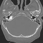
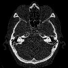
CT
High resolution bony CT reformats is the modality of choice for assessing the external acoustic canal. A number or key points should be looked for and specifically mentioned in reports as it impacts on surgical reconstruction.
- middle ear cavity volume: a width of greater than 3 mm is usually needed for successful surgery
- ossicles
- malleus: usually has a rudimentary handle (as there is no normal tympanic membrane)
- incudomalleolar joint: usually normal
- incus: usually normal
- stapes: important to note as an abnormal or absent stapes needs to be replaced with a prosthetic
- inner ear structure
- both the oval and round window need to be present for successful surgery
- course of internal carotid artery, and location of the jugular bulb: if abnormal can be hazardous during surgery
- course of facial nerve: often abnormally anterior and can be damaged during reconstruction
Treatment and prognosis
Surgical reconstruction requires formation of a new EAC and new tympanic membrane (usually with temporalis fascia). The ossicles often need to be mobilized.
Differential diagnosis
- EAC exostosis (surfer's ear)
Siehe auch:
- Arteria carotis interna
- Malleus
- Trommelfell
- Crouzon-Syndrom
- Franceschetti-Zwahlen-Syndrom
- congenital cholesteatoma
- Amboss
- Goldenhar-Gorlin-Syndrom
- Meatus acusticus externus
- Innenohr
- Pierre-Robin-Sequenz
- Ohrmuschelfehlbildung
- Fehlbildungen der Gehörknöchelchen
- Stenose des äußeren Gehörgangs
- external ear
und weiter:


 Assoziationen und Differentialdiagnosen zu Gehörgangsatresie: