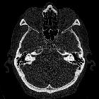
inner ear

This image
is part of a series which can be scrolled interactively with the mousewheel or mouse dragging. This is done by using Template:Imagestack. The series is found in the category Temporal bone in computertomography case 001. Felsenbein in der Computertomographie. Bilder für scrollbaren Stapel.

Middle ear
• Inner ear (illustrations) - Ganzer Fall bei Radiopaedia

The cochlea
and vestibule, viewed from above. All the hard parts which form the roof of the internal ear have been removed with the saw.


Position of
the right bony labyrinth of the ear in the skull, viewed from above. The temporal bone is considered transparent and the labyrinth drawn in from a corrosion preparation. (Spalteholz.)


Inner ear •
Inner and middle ear anatomy - Ganzer Fall bei Radiopaedia
The inner ear refers to the bony labyrinth, the membranous labyrinth and their contents. It may also be referred to as the vestibulocochlear organ, supplied by the vestibulocochlear nerve (CN VIII). It is divided into three main parts:
- the cochlea housing the cochlear duct for hearing
- the vestibule housing the utricle and saccule for static balance
- the semicircular canals housing the semicircular ducts for kinetic balance
As the membranous labyrinth is slightly smaller than the osseous labyrinth, the two are separated by perilymph, which does not communicate with the endolymph contained in the membranous labyrinth.
Siehe auch:
- Cavum tympani
- Cochlea
- Felsenbein
- Mondini-Dysplasie
- Labyrinthitis
- Tumor des Saccus endolymphaticus
- fenestrale Otosklerose
- ossifizierende Labyrinthitis
- Michel-Aplasie
- erweiterter Aquaeductus vestibuli
und weiter:

 Assoziationen und Differentialdiagnosen zu Innenohr:


