Patellaluxation

Patellaluxation
und Reposition - kleine Abscherung aus der medialen Facette. Typische Kontusion auch an der Anschlagstelle am lateralen Femurkondylus.

Patella alta
• Transient lateral patellar dislocation - Ganzer Fall bei Radiopaedia

Lateral
patellar dislocation • Lateral patellar dislocation - Ganzer Fall bei Radiopaedia

Lateral
patellar dislocation • Transient lateral patellar dislocation - Ganzer Fall bei Radiopaedia

Lateral
patellar dislocation • Lateral patellar dislocation - Ganzer Fall bei Radiopaedia

MRI after
luxation of the patella (axial). There is bone bruise at the medial surface of the patella itself and in the corresponding surface of the lateral condyle of the femur (arrow). The medial retinaculum of the patella is disrupted.

MRI after
luxation of the right patella. There is bone bruise at the medial surface of the patella (axial image) and in the corresponding surface of the lateral condyle of the femur (coronal). The medial retinaculum of the patella is at least partially disrupted.

X-ray and MRI
after luxation of the patella. There is a fragment and bone bruise at the medial surface of the patella and in the corresponding surface of the lateral condyle of the femur. The medial retinaculum of the patella is disrupted.


Patellaluxation
im Röntgenbild. Rechtes Knie, tangentiale Aufnahme. Der Femurkondylus ist nicht komplett mit abgebildet.


Patellaluxation
im Röntgenbild a.p. Linkes Knie, Luxation wie meistens nach lateral (aussen).

School ager
with knee pain. AP radiographs of the knees show lateral patellar dislocation bilaterally along with bilateral gracile bones in the femurs, tibiae and fibulae.The diagnosis was Trisomy 18.

Chronische
Patellaluxation bei Tetraspastik seit der Kindheit. Die Patella findet sich lateral des Femurcondylus. Dadurch ist auch die Tibia deutlich pathologisch nach außen rotiert.

Lateral
patellar dislocation • Illustration for lateral patellar dislocation - Ganzer Fall bei Radiopaedia

Lateral
patellar dislocation • Lateral patellar dislocation - Ganzer Fall bei Radiopaedia

Lateral
patellar dislocation • Recent patella dislocation (MRI) - Ganzer Fall bei Radiopaedia

Radiograph of
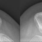
lateral luxation of the patella. Left before treatment, right after reduction. Even after reduction, the patella is still displaced to the outside.

Lateral
patellar dislocation • Lateral patellar dislocation - Ganzer Fall bei Radiopaedia

Lateral
patellar dislocation • Lateral patellar dislocation - Ganzer Fall bei Radiopaedia

Lateral
patellar dislocation • Patellar dislocation - Ganzer Fall bei Radiopaedia

Lateral
patellar dislocation • Lateral patellar dislocation - Ganzer Fall bei Radiopaedia

Lateral
patellar dislocation • Transient lateral patellar dislocation relocation - Ganzer Fall bei Radiopaedia

Lateral
patellar dislocation • Medial femoral condyle fracture associated with lipohemarthrosis and lateral patellar dislocation - Ganzer Fall bei Radiopaedia

Lateral
patellar dislocation • Patellar dislocation - Ganzer Fall bei Radiopaedia

Lateral
patellar dislocation • Lateral patellar dislocation - Ganzer Fall bei Radiopaedia

Lateral
patellar dislocation • Lateral patellar dislocation - Ganzer Fall bei Radiopaedia

Lateral
patellar dislocation • Lateral patellar dislocation with avulsion fracture - Ganzer Fall bei Radiopaedia

Lateral
patellar dislocation • Transient lateral patellar dislocation with osteochondral injury - Ganzer Fall bei Radiopaedia

Intra-articular
loose bodies • Patellar dislocation with chondral injury - Ganzer Fall bei Radiopaedia

Teenager who
fell on their right knee and has pain. AP (above) and Merchant (below left) radiographs of the right knee show lateral displacement of the patella and a small avulsion fragment (sliver sign) medial to the patella. Merchant radiograph of the right knee post reduction (below right) shows appropriate reduction of the patella along with medial soft tissue swelling.The diagnosis was lateral patellar dislocation.
Patellaluxation
Siehe auch:
- Jägerhutpatella
- Patelladysplasie
- mediales Retinaculum patellae
- Operation nach Blauth (Patellasehne)
- transient lateral patellar dislocation
- Scholte-Syndrom
- Ruptur mediales Retinaculum
- patella fracture-dislocation
- patellar subluxation
- Roux-Goldthwait procedure
- Prieto-Syndrom
- Patellaluxation bei Trisomie 18
- chronische Patellaluxation
und weiter:

 Assoziationen und Differentialdiagnosen zu Patellaluxation:
transient
lateral patellar dislocation


