Beckenasymmetrie
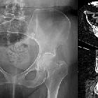
Rechtsseitige
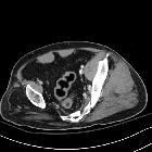
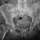
Beckenatrophie bei Poliomyelitis in der Kindheit: In der Computertomografie rechts (oben axial, unten koronar) zeigt sich die massive fettige Atrophie der Beckenmuskulatur rechts. Im Röntgenbild sieht man die deutliche Hypoplasie mit Coxa valga. (In der axialen Computertomografie mitdargestelltes Stoma im rechten Unterbauch.)
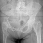
Poliomyelitis
• Polio pelvis - Ganzer Fall bei Radiopaedia
Poliomyelitis
• Polio - Ganzer Fall bei Radiopaedia

Proximal
femoral focal deficiency • Proximal focal femoral deficiency - Ganzer Fall bei Radiopaedia

Developmental
dysplasia of the hip • Chronic hip dysplasia - Ganzer Fall bei Radiopaedia
Developmental
dysplasia of the hip • Chronic hip dysplasia - Ganzer Fall bei Radiopaedia

Developmental
dysplasia of the hip • Chronic hip dysplasia - Ganzer Fall bei Radiopaedia

Developmental
dysplasia of the hip • Unilateral hip dysplasia with dislocation and pseudoarticulation - Ganzer Fall bei Radiopaedia

Developmental
dysplasia of the hip • Untreated right sided developmental dysplasia of the hip - Ganzer Fall bei Radiopaedia

 Assoziationen und Differentialdiagnosen zu Beckenasymmetrie:
