Proximaler Femurdefekt
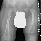


Proximal femoral focal deficiency is a congenital partial absence of the proximal end of the femur with shortening of the entire lower limb. The diagnosis and classification have been based mainly on plain radiograph findings. This method does not permit definite classification during the first year of life.
Epidemiology
The incidence of proximal focal femoral deficiency is on the order of 1-2 per 100,000 live births .
Pathology
The etiology of this disorder is uncertain. There may be an association with in utero exposure to thalidomide . In severe cases, the proximal femur, femoral head and neck, and acetabulum are absent.
Associations
- fibular hemimelia
- absent patella
Classification
A number of classifications have been proposed, with the Aitken four class (A-D) (modified by Amstutz) most commonly used. See classification of proximal focal femoral deficiency.
Radiographic features
MRI
MR imaging is used to define the cartilaginous proximal femur and the presence or absence of a cartilaginous connection to the femoral head. Therapeutic decisions are based on the detection of a femoral head and the presence of a connection.
The severity of coxa vara, if present, also influences treatment. GRE imaging to clearly depict cartilage is of particular value. Routine coronal and axial MR images may be adequate with oblique images required in some cases.
MR arthrography
Arthrography is useful for confirming the presence or absence of a femoral head and may be used as an adjunct to standard non-contrast MRI.
Treatment
Treatment depends on the degree of deficiency and clinical presentation. For mild cases, reconstruction of the hip with femoral or pelvic osteotomies is possible . In severe cases, complications associated with surgical correction may outweigh the potential for benefit and alternative treatments such as rotationplasty or amputation may be considered .
Siehe auch:
und weiter:


 Assoziationen und Differentialdiagnosen zu Proximaler Femurdefekt: