Femur


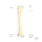


The femur (plural: femora) is the longest, most voluminous and strongest bone in the human body.
Gross anatomy
It is composed of the upper extremity, body and lower extremity and provides several muscular origins and insertions.
Proximal portion
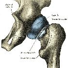
The upper extremity is composed of the head, neck, greater trochanter and lesser trochanter. The head of the femur articulates with the acetabulum of the pelvis to create the hip joint.
Femoral head and neck
The femoral head is supported by the neck of the femur. It is globular and forms rather more than a hemisphere. It is directed caudally, medially and anteriorly. Its surface is smooth and coated in cartilage except for an ovoid depression (the fovea capitis femoris) which is situated a little below and behind the center of the head. The fovea allows for the attachment of the ligament of head of the femur.
If there is a fracture of the neck of femur, the blood supply through the ligament becomes crucial. Where this occurs, there is a risk of insufficient blood supply to the head and avascular necrosis may occur. It is for this reason that the treatment of choice is a total hip replacement rather than dynamic hip screwing.
Greater and lesser trochanters
The greater trochanter is a large, irregular, quadrilateral eminence attached to the proximal end of the femur. It is directed laterally and posteriorly and, in the adult, is about 1 cm lower than the femoral head. It has two surfaces and four borders to which several muscles are attached.
The lesser trochanter is a conical eminence which is of variable size. It projects from the posterior surface of the neck of the femur. It is continuous with the middle division of the linea aspera (the prominent ridge along the femur) and the intertrochanteric crest. Like the greater trochanter, it is also involved in muscular attachment.
Muscles
- gluteus medius
- gluteus minimus
- piriformis
- obturator internus
- gemelli
- obturator externus
- quadratus femoris
- vastus lateralis
- iliopsoas
Femoral shaft
The femoral shaft is almost cylindrical in form, is a little broader superiorly and is slightly arched such that it is convex anteriorly and concave posteriorly where it is strengthened by a prominent longitudinal ridge of bone, the linea aspera. A variety of muscles have their origins at and insert into the femoral shaft (all, apart from gluteus maximus and vastus intermedius, interact with the posterior surface of the bone):
- gluteus maximus
- pectineus
- adductor longus
- adductor brevis
- adductor magnus
- adductor minimus
- vastus medialis
- vastus lateralis
- vastus intermedius
- biceps femoris
- articularis genu
Distal portion
The lower extremity is composed of the medial and lateral condyles, adductor tubercle, patellar surface and intercondylar fossa. It is larger than the upper extremity and articulates with the tibia and patella at the knee.
Anteriorly, the condyles are slightly prominent and separated from one another by the patellar surface. Posteriorly, they project considerably and the interval between them forms a deep notch, the intercondylar fossa. The lateral condyle is more prominent than the medial.
The articular surface of the lower end of the femur occupies the anterior, inferior and posterior surfaces of the condyles.
A number of muscles originate from the distal femur:
Development
The femur ossifies in cartilage. A center in the shaft appears in utero (8 week) and a center at distal femur appears at birth (end of 9 fetal month) with its presence a medicolegal evidence of maturity. The distal portion is the growing end with its epiphysis uniting with the shaft after 20 years.
A center appears in the femoral head at the age of 1, greater trochanter at 3, and lesser trochanter at 12 years. These fuse with the shaft at about 18 years of age.
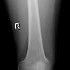
Relevant pathology
Siehe auch:
und weiter:


 Assoziationen und Differentialdiagnosen zu das Femur:
