Fibulare Hemimelie

Fibular hemimelia is a congenital lower limb anomaly characterized by partial or complete absence of the fibula and includes a spectrum ranging from mild fibular hypoplasia to complete fibular aplasia .
Epidemiology
Although rare in occurrence, it is the most common congenital absence of long bone of the extremities . The incidence has been suggested to be approximately 5.7 to 20 cases per 1 million births . This condition is twice as common in boys as in girls.
Presentation
Fibular hemimelia is usually obvious at birth with limb shortening and limb-length discrepancy . Syndactyly, oligodactyly or polydactyly may also be present . It may also be detected antenatally during obstetric ultrasound evaluation for fetal anomalies .
Pathology
Several theories have been put forward to explain this condition like defects in the apical ectoderm ridge, defects secondary to an absent anterior tibial artery . Amniotic bands may be one of the causal factors to insult to the growing limb bud in utero resulting in this condition . Kumar D and Krishnamoorthy S et al. reported a case of congenital absence of femur and fibular hemimelia related to maternal hyperpyrexia .
Associations
Fibular hemimelia is usually an isolated anomaly and occurs sporadically. However, it can be associated with proximal focal femoral deficiency, absence of lateral rays and phalanges of lateral toes , syndactyly and polydactyly .
Classification
One of the commonly used classifications for fibular hemimelia is that of Achterman and Kalamachi et al. which divides the condition into two types:
- type I: minimal hypoplasia of the fibula
- type II: complete absence of the fibula
Radiolographic features
Fetal sonography
- unilateral short-for-date lower limb
- non-visualization of two bones in the leg region
- associated short femur may be seen in cases of proximal femoral focal deficiency
- talipes equinovalgus deformity of the affected foot
- syndactyly involving lateral toes mainly the fourth and fifth toes
Plain radiograph
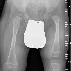
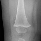
- absence of the fibula, needs to be seen in both frontal and lateral radiographs as the fibula; may be masked by the tibia in one view
- shortening of the femur
- lateral ray malformations
CT
- absence of the fibula
- absence or malformations of the lateral ray
- syndactyly and polydactyly as the case may be
- 3D study is required for assessment of limb length discrepancies and planning of surgical management
MRI
- same features as can be seen with plain radiographs and CT imaging
- additional pseudoarthrosis of the femur may be assessed in cases of proximal femoral focal deficiency
- assessment of muscle bulk and agenesis
Management
The management is mainly surgical and includes limb-lengthening procedures like Ilizarov's technique or amputation with a prosthesis. Residual deformity and disability is more usual than not.
Siehe auch:
und weiter:


 Assoziationen und Differentialdiagnosen zu Fibulare Hemimelie: