ANCA-assoziierte Vaskulitiden

Imaging of
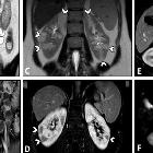
intestinal vasculitis focusing on MR and CT enterography: a two-way street between radiologic findings and clinical data. Intestinal vasculitis in a 39-year-old female presenting with episodic abdominal pain and findings of non-specific terminal ileitis in ileocolonoscopy examination. MRE was obtained. Coronal T2-W and post-contrast T1-W images (A, B) display mild mural thickening and hyperenhancement at distal terminal ileum (thick white arrows). Coronal T2-W and post-contrast T1-W images (C, D) reveal multiple peripheral wedge-shaped hyperintense areas at both kidneys, showing decreased enhancement on delayed phase (white arrowheads). Axial post-contrast T1-W and corresponding DWI images (E, F) show a hypoenhancing lesion at the right renal cortex with diffusion restriction (thin white arrows). Vasculitis was suspected as the underlying cause. Afterward, positive results for antineutrophil cytoplasmic antibody (ANCA) and inflammatory markers were consistent with the diagnosis of ANCA-associated vasculitis
Anti-neutrophil cytoplasmic antibody (ANCA) - associated vasculitides refer to a group of heterogeneous autoimmune diseases characterized by necrotizing vasculitides and positive ANCA titers. They are reactive to either proteinase-3 (PR3-ANCA) - cANCA or myeloperoxidase (MPO-ANCA) - pANCA. These vasculitides affect arterioles, capillaries, and venules which can involve virtually any organ with varying degrees of severity. As a general rule, the lungs and kidneys are the most commonly involved.
The conditions that fall into this group are:
- granulomatosis with polyangiitis
- positive to cANCA in about 90% of cases
- granulomatous
- microscopic polyangiitis
- positive to pANCA in about 50-90% of cases
- non-granulomatous
- idiopathic pauci-immune pulmonary capillaritis
- sometimes classified as a subset of microscopic polyangiitis
- may or may not be positive to pANCA
- non-granulomatous
- idiopathic pauci-immune rapidly progressive glomerulonephritis (RPGN)
- sometimes classified as a subset of microscopic polyangiitis
- may or may not be positive to pANCA
- non-granulomatous
- eosinophilic granulomatosis with polyangiitis
- positive to pANCA in about 35-75% of cases
- granulomatous
See also
Siehe auch:
und weiter:

 Assoziationen und Differentialdiagnosen zu ANCA-assoziierte Vaskulitiden:
